Category: Understanding the tests

  • Dr. Gurpreet Singh Padda presenting beside the title card The Normal B12 That Wasn't, The Angry Gut, Chapter 6

    Atrophic Gastritis: When Your Stomach Stops Absorbing B12

    The Normal B12 That Wasn't | The Angry Gut, Chapter 6

    Atrophic Gastritis: When Your Stomach Stops Absorbing B12

    In atrophic gastritis the stomach lining loses its parietal cells, the only source of intrinsic factor, the protein that escorts vitamin B12 into the body. With fewer of those cells, B12 travels through without getting in, and a normal blood B12 does not clear the stomach.

    A stomach lining can lose the cells that capture vitamin B12 long before any blood count changes. Here is what that means for your numbers, and which measurements can see the damage.

    Atrophic gastritis is a thinned, worn-down stomach lining, and it may be present in up to 15% of American adults. Most of them never hear the words, because the condition rarely starts with stomach pain. It shows up years later and far from the stomach: numb feet, a slipping memory, an iron level nobody could explain. The video The Normal B12 That Wasn’t, from The Angry Gut by Dr. Gurpreet Singh Padda, MD, MBA, MHP, and Ami Michelle Grimes, follows one man through four lost years. The lens here is measurement: what a failing lining does to your lab results, what a routine visit checks, and what it leaves unmeasured.

    What atrophic gastritis takes away

    The gut is the first brain. It decides what enters the body, and the brain in the skull, the second brain, lives on whatever it lets through. Vitamin B12 has the most demanding entry process of any nutrient. Acid pries it loose from food. A protein called intrinsic factor, made only by the parietal cells of the stomach lining, escorts it down the small bowel to a receptor at the far end. In a failing lining those parietal cells are being lost. Fewer cells means less escort, and the vitamin travels through without getting in.

    Two biological forces do most of the damage. One is infection: people carrying H. pylori develop new atrophy at 5.0 times the rate of people who are not infected. The other is autoimmunity, the immune system attacking the parietal cells directly. The infection is receding worldwide; the autoimmune form is not. A third force is economic. We subsidize the calories that feed type 2 diabetes and reflux, then treat both with long-term drugs that also pull B12 down, so the lining and the prescriptions push in the same direction. The background of low-grade metabolic inflammation, metaflammation, is the terrain that stomach lives in.

    Why a normal B12 does not clear your stomach

    The measurement gap fits in one sentence: nobody measures absorption anymore. The Schilling test once did, and nothing has replaced it. What is left is a blood level, which answers a smaller question. The man in the video had a B12 of 348, was told it was fine, and had almost no parietal cells left. Why a lone serum B12 misleads, and how a functional marker fills the hole, is covered in the methylmalonic acid test explained. For the stomach the lesson is different. Even a correctly low B12 is a late signal. By the time the vitamin runs short, the lining has usually been failing for a long while.

    What are the early signs of atrophic gastritis?

    A failing lining rarely runs short of one thing. Iron tends to go first. When atrophy sits in the acid-making body of the stomach, iron deficiency appears in as many as half of patients, often well before B12 falls, because iron stores drain faster. An old low ferritin that was written off years ago can be the first footprint.

    Symptoms are a weak guide. Pooled across many studies, these stomach changes were not statistically more common in people with complaints than in people without them, so waiting for heartburn means waiting too long. The better clues are the company the disease keeps. Autoimmune thyroid disease is found in 36% to 44% of people with autoimmune gastritis, and type 1 diabetes carries roughly three to five times the general population’s risk.

    The textbook picture of who gets it is also off. In a Miami biopsy series drawn heavily from Hispanic patients, autoimmune gastritis was found in 4.6%, compared with 1.1% in a Baltimore series of mostly White patients. Being younger, male, Hispanic or Black does not take it off the table.

    Which blood tests point to atrophic gastritis?

    Two antibodies look for the autoimmune attack, and each one fails in its own direction. Anti-intrinsic factor antibody was 100% specific in one assay comparison but caught only 38% of cases, and it tends to turn positive late. Anti-parietal cell antibody catches more but proves less, and it grows less reliable from age 50 on. Ordered together, the pair reached a sensitivity of 90% and a specificity of 95.7%. If only one was drawn and it was negative, the question is still open.

    Two more traps are worth knowing about. Pepsinogen testing, used to estimate atrophy in some countries, is not available for routine clinical use in the United States. And gastrin, another marker used to locate atrophy, is pushed up three to five times by acid-blocking drugs, so it can end up reporting on the pill rather than the stomach. Planning around that is your physician’s call; do not stop a medication on your own.

    No blood test is the last word. The lining itself is examined by a gastroenterologist with a scope and biopsies, a different test done elsewhere. In one recent study, only 9% of people diagnosed with pernicious anemia were offered a gastroscopy at the time of diagnosis.

    What Measura shows in the same person

    Measura [Cardiometabolic and Autonomic Health Analysis] does not scope stomachs and does not diagnose gastritis. It measures what a starved nerve and a strained metabolism leave behind, which is exactly the part a stomach workup never looks at. Results go to your physician, who reads them against the rest of your chart.

    • Small nerves. Sudomotor testing looks at sweat-gland function in the small nerve fibers of the hands and feet, the fibers that often complain first as burning or numbness.
    • Balance. Vestibular and balance testing puts a number on unsteadiness, which matters once numb feet start changing how someone walks.
    • Memory. A cognitive assessment sets a baseline, so a later change is measured instead of guessed.
    • Metabolism. Laboratory panels map the insulin resistance and inflammation that so often put metformin and an acid blocker in the medicine cabinet to begin with.

    Together those numbers answer what the stomach tests cannot: how much the shortfall has already cost the nerves and the brain, and whether that picture improves once the cause is dealt with.

    What to ask at your next visit

    For years I told patients that stomach acid simply fades as we get older. The measurements say I was wrong. Most older stomachs still make acid, and the ones that stop are usually diseased, not merely old. That correction changes the question to bring. Not whether you are just aging, but whether your stomach lining still works.

    • Has anyone drawn a methylmalonic acid or holotranscobalamin, or only a total B12?
    • Was my iron ever low, even years ago?
    • Were both stomach antibodies checked, or only one?
    • Do I have thyroid autoimmunity or type 1 diabetes that raises the odds?
    • How long have I taken metformin or an acid blocker?
    • Has the stomach lining itself ever been examined?

    If the person losing ground is an aging parent, B12 deficiency in older adults covers the family side. Every study behind these numbers sits in the companion deep dive for The Angry Gut. Physicians weighing the screening side can read the clinical version for practices.

    Frequently asked questions

    What does atrophic gastritis mean on a biopsy report?

    It means the glands of the stomach lining have thinned and some have been lost, usually from long-term H. pylori infection or an autoimmune attack. Those glands include the cells that make acid and intrinsic factor, so B12 and iron absorption can suffer. It is a precancerous condition, but progression to stomach cancer is uncommon, about 0.1% to 0.3% per year. Read what a test result can and cannot tell you.

    Can atrophic gastritis cause numbness in the feet?

    It can, by an indirect route. When the lining stops making enough intrinsic factor, B12 stops getting in, and the long nerves of the legs are among the tissues that suffer over years. Numbness and burning have many other causes, diabetes among them, so the pattern needs a physician’s workup rather than a guess. See what numbness, burning and tingling can mean.

    Are B12 shots better than pills if my stomach is damaged?

    It depends on the cause, and the choice belongs to your physician. About 1% of an oral dose crosses the gut wall without any intrinsic factor, which is why very high-dose tablets can work for some people. British guidance still advises lifelong injections when autoimmune gastritis is the cause. Bring these questions worth asking your doctor.

    Can Measura test for atrophic gastritis?

    No. It is confirmed by a gastroenterologist with a scope and biopsies, which is a different test done elsewhere. Measura measures what a long shortfall may already be doing downstream: small-fiber nerve function, balance, cognition and the metabolic picture. Those results go to your physician to sit beside the stomach workup. Learn what Measura actually measures.

    Does taking an acid blocker for years affect B12?

    Long use is linked to lower B12. In a large study, two or more years of proton pump inhibitor supply carried an odds ratio of 1.65 for B12 deficiency, and higher daily doses carried more. Most deficient people in that study were not on the drug, and the drug has real uses, so any change is a conversation with your physician. See how to approach understanding your results.

    Is atrophic gastritis serious?

    It can be, mostly because of what it quietly takes away. A failing lining can starve the nerves and brain of B12 for years, showing up as numb feet, a slipping memory or unexplained low iron long before any stomach pain. It is also a precancerous condition, although progression to stomach cancer is uncommon, about 0.1% to 0.3% per year. Waiting for symptoms means waiting too long.

    What causes atrophic gastritis?

    Two forces do most of the damage. One is H. pylori infection: people carrying it develop new atrophy at 5.0 times the rate of people who are not infected. The other is autoimmunity, the immune system attacking the parietal cells of the stomach lining directly. The infection is receding worldwide; the autoimmune form is not. Either way, the cells that make intrinsic factor are lost.

    How is atrophic gastritis diagnosed?

    A gastroenterologist confirms it with a scope and biopsies of the stomach lining. Blood tests point toward it: anti-intrinsic factor antibody and anti-parietal cell antibody ordered together reached 90% sensitivity and 95.7% specificity, while either one alone misses cases. A normal total B12 does not rule it out. In one recent study, only 9% of people diagnosed with pernicious anemia were offered a scope at diagnosis.

    See what the shortfall has already touched

    Ask about nerve, balance, cognitive and metabolic testing so your physician can see the downstream picture beside the stomach workup.

    4477 Woodson Rd, Suite 201, St. Louis, MO 63134. Monday to Friday, 9:00 a.m. to 5:00 p.m. Please do not send symptoms, diagnoses or images through a web form — a website form is not a secure medical channel. Send your name and number and we will call you back.

    References

    • Shah, S. C., Piazuelo, M. B., Kuipers, E. J., & Li, D. (2021). AGA Clinical Practice Update on the Diagnosis and Management of Atrophic Gastritis: Expert Review. Gastroenterology, 161(4), 1325–1332.e7. https://doi.org/10.1053/j.gastro.2021.06.078
    • Osmola, M., Hémont, C., Romańczyk, M., Druet, A., Chapelle, N., Matysiak-Budnik, T., Lenti, M. V., & Martin, J. C. (2025). A comparative study of different assays for autoantibodies detection in patients with autoimmune gastritis. Journal of Translational Autoimmunity, 10, 100294. https://doi.org/10.1016/j.jtauto.2025.100294
    • Poveda, J. C., Park, J. Y., Garcia-Buitrago, M. T., Singhi, A., Alruwaii, Z., Kumar, S., McDonald, O. G., & Montgomery, E. A. (2024). Autoimmune Metaplastic Atrophic Gastritis (AMAG): Regional Demographics and Their Effect on Prevalence. International Journal of Surgical Pathology, 33(3), 565–570. https://doi.org/10.1177/10668969241271311
    • Guéant, J.-L., Guéant-Rodriguez, R.-M., & Alpers, D. H. (2022). Vitamin B12 absorption and malabsorption. Vitamins and Hormones, 119, 241–274. https://doi.org/10.1016/bs.vh.2022.01.016
    • Lam, J. R., Schneider, J. L., Zhao, W., & Corley, D. A. (2013). Proton pump inhibitor and histamine 2 receptor antagonist use and vitamin B12 deficiency. JAMA, 310(22), 2435–2442. https://doi.org/10.1001/jama.2013.280490
    • Feldman, M., Cryer, B., McArthur, K. E., Huet, B. A., & Lee, E. (1996). Effects of aging and gastritis on gastric acid and pepsin secretion in humans: a prospective study. Gastroenterology, 110(4), 1043–1052. https://doi.org/10.1053/gast.1996.v110.pm8612992
    • Rustgi, S. D., Bijlani, P., & Shah, S. C. (2021). Autoimmune gastritis, with or without pernicious anemia: epidemiology, risk factors, and clinical management. Therapeutic Advances in Gastroenterology, 14, 17562848211038771. https://doi.org/10.1177/17562848211038771
    • Thain, A., Hart, K., & Ahmadi, K. R. (2025). Addressing the gaps in the vitamin B12 deficiency 2024 NICE guidelines: Highlighting the need for better recognition, diagnosis, and management of pernicious anaemia. European Journal of Clinical Nutrition, 79(7), 607-610. https://doi.org/10.1038/s41430-025-01583-4
    • Mülder, D. T., Hahn, A. I., Huang, R. J., Zhou, M. J., Blake, B., Omofuma, O., Murphy, J. D., Gutiérrez-Torres, D. S., Zauber, A. G., O’Mahony, J. F., Camargo, M. C., Ladabaum, U., Yeh, J. M., Hur, C., Lansdorp-Vogelaar, I., Meester, R., & Laszkowska, M. (2024). Prevalence of Gastric Precursor Lesions in Countries With Differential Gastric Cancer Burden: A Systematic Review and Meta-analysis. Clinical Gastroenterology and Hepatology, 22(8), 1605-1617.e46. https://doi.org/10.1016/j.cgh.2024.02.023
    • Chan, C. Q. H., Low, L. L., & Lee, K. H. (2016). Oral vitamin B12 replacement for the treatment of pernicious anemia. Frontiers in Medicine, 3, 38. https://doi.org/10.3389/fmed.2016.00038


    Related reading

  • Dr. Gurpreet Singh Padda in a white coat beside the title card reading Chapter 4, The Angry Gut, The Permeable Fortress

    How to Test for Leaky Gut, and Why Zonulin Is the Wrong Test

    The Leaky Gut Test You Paid For Cannot See the Wall | The Angry Gut, Chapter 4

    How to Test for Leaky Gut, and Why Zonulin Is the Wrong Test

    Leaky gut is tested with a physician-ordered sugar permeability test: you drink sugars of two sizes, then collect urine over timed windows, with labeled mannitol and each sugar read separately. The mail-order zonulin blood kit is the wrong test.

    A zonulin number on a printout feels like an answer. It is not a measurement of your gut wall, and the harm that follows a leak shows up in numbers most people never get.

    If you are trying to work out how to test for leaky gut, the zonulin test is probably the first thing you found. It arrives by mail, it prints one number, and people rebuild their whole diet around it. Chapter 4 of The Angry Gut by Dr. Gurpreet Singh Padda, MD, MBA, MHP, and Ami Michelle Grimes, told in the video above, lands somewhere sharper than either side of that fight: the wall is real, its failure can be measured, and the kit most people order does not measure it.

    Dr. Padda is candid that his training taught him to wave these printouts away. He dropped the habit after reading a study in which a leaking wall, measured properly in healthy people, showed up years before disease did.

    What a zonulin result is really reporting

    Picture the lining of your gut as a wall one cell thick. It is the outer surface of the first brain, the gut, which stays in constant conversation with the second brain inside your skull. The mortar between its cells is sealed by proteins called claudins. When that seal loosens, fragments of gut bacteria slip into the blood, and metaflammation, the low and steady metabolic inflammation behind so much chronic disease, has a place to start.

    Zonulin was meant to be a blood readout of that seal. The trouble is the assay. Zonulin was defined as a single protein, pre-haptoglobin-2, which some people’s genes do not let them make. When researchers compared the most widely used commercial kit with each person’s genotype, the kit reported the protein in people who cannot produce it, and it failed to detect the purified protein at all. What it seems to bind is properdin, an unrelated immune protein. A second research group ran two other commercial kits and found haptoglobin and a complement protein on the other end of the antibody.

    The decisive comparison put serum zonulin next to a direct permeability test in the same people, healthy relatives of patients with Crohn’s disease. The correlation was r2 = 0.004.

    How to test for leaky gut: what a real permeability test involves

    You test a wall by seeing what gets across. For a permeability test, you drink sugars of two different sizes, then collect urine during set time windows. A small sugar slips through a healthy lining; a large one mostly should not. The early window reflects the small bowel and the late window reflects the colon. A physician orders it, and it is a timed urine collection rather than a blood kit.

    Three details decide whether it can be trusted:

    • Labeled mannitol. Everyday mannitol comes from food. In the study that validated the test, it was already in the urine of all 60 healthy volunteers before anyone swallowed a dose.
    • Each sugar read separately. In people with inflammatory bowel disease, the two sugars measured one at a time separated patients from healthy volunteers. The ratio between them, the figure most reports print, found no difference.
    • A second look before any verdict. People differ enormously on this test. In one group of 39 healthy young adults, 14 landed on the abnormal side of a common cutoff.

    Measured this way, the result carries weight. Healthy relatives of people with Crohn’s disease, 1,420 of them, were tested and then followed for a median of 7.8 years. An abnormal baseline result came with roughly triple the risk of a later Crohn’s diagnosis, a hazard ratio of 3.03. That group carried strong family risk for one disease. A leaking wall is a finding worth acting on, not a sentence.

    What can cause a leaky gut in an ordinary week?

    The exposures shown to loosen the human gut lining are mostly things people do on purpose. One anti-inflammatory tablet more than doubled small-bowel leakiness in people with healthy guts, and a public speech raised it as well. One evening of heavy drinking pushed blood endotoxin up within 30 minutes. Raising core temperature two degrees, with no exercise, opened the small bowel in younger and older adults alike.

    Behind that list sits a system problem. A bottle of anti-inflammatories on the shelf is quicker to reach than a workup for why the pain keeps returning, and short, crowded appointments reward the refill over the question. If you take one every day, do not stop on your own. Ask your physician what it is covering and whether that cause has ever been traced.

    Is a leaky gut dangerous?

    Endotoxin is a piece of the outer coat of certain gut bacteria, and your immune system reads it as an invasion. When healthy volunteers were given a small intravenous dose, their insulin sensitivity dropped 35% within a day, even though the pancreas kept producing insulin normally. Over ten years in 7,169 adults, people in the top quarter of endotoxin activity had 52% more risk of developing diabetes than people in the bottom quarter.

    Here is the detail most leaky gut advice skips. After a 910-calorie fast-food meal, endotoxin in the blood rose and the inflammatory switches inside white blood cells turned on. C-reactive protein, the inflammation test most clinics order, did not budge.

    Body size is a poor guide as well. In a study of women in The Gambia that measured endotoxin by mass spectrometry, levels were 57% higher in women with obesity and diabetes, yet no different between women with obesity and women without it. The gradient followed diabetes, not weight. The numbers worth having after a leak are metabolic, not the bathroom scale.

    What Measura measures here, and what it does not

    Measura [Cardiometabolic and Autonomic Health Analysis] does not offer zonulin or intestinal permeability testing. If your physician decides a permeability test is worth doing, the timed sugar collection is arranged elsewhere, with labeled mannitol.

    What Measura can measure is the terrain a leaking wall acts on:

    Every result goes to your physician. Measura measures; it does not diagnose leaky gut or treat it.

    Questions to bring to your physician

    • Is a permeability test reasonable for me, and would it use labeled mannitol?
    • Would the report show each sugar separately, not only the ratio?
    • If my result sits near the cutoff, can we repeat it before I change what I eat?
    • What would we do differently because of the zonulin number I already have?
    • Can we measure my insulin sensitivity and body composition to see whether anything that crossed has left a mark?

    How the first brain signals the second brain is traced in the previous post on the vagus nerve, and everything that crosses the wall reaches the liver first, which is where the next post on an echogenic liver picks up. Every study above, including the results that cut against the argument, is laid out in the Chapter 4 book companion. Unmeasured is unmanaged: a wall can be closed, and what it let through can be tracked.

    Frequently asked questions

    Is the zonulin test accurate for leaky gut?

    Not as a measure of the gut wall. Kits sold as zonulin tests failed validation in two separate laboratories, and neither check found the protein printed on the label. When serum zonulin and a direct sugar test were run side by side in the same people, the two did not correlate. Why one number rarely settles a question is covered in what a test result can and cannot tell you.

    What does a high zonulin level mean?

    It means the kit detected something that tends to rise with obesity, diabetes, and glucose and lipid markers. It does not mean your gut wall is breached, and it is not a reason on its own to remove foods. If metabolic trouble is what the number hints at, measuring insulin and body composition answers that directly. For reading any flagged value, see understanding your results.

    Can a healthy person get an abnormal leaky gut result?

    Yes. People vary widely on sugar permeability tests, and the usual cutoffs sit close to normal. In one group of healthy young adults, more than a third landed on the abnormal side of the line. A borderline result calls for a repeat under controlled conditions before any change in diet. How repeat testing is usually planned is explained in how often to repeat testing.

    Does Measura test for leaky gut?

    No. Zonulin and intestinal permeability tests are not part of the Measura library; a permeability test is a timed urine collection your physician arranges elsewhere. Measura measures the metabolic and body composition picture a leaking wall can affect, and every finding goes to your physician. For the full list, read what Measura actually measures.

    Can a leaky gut cause insulin resistance?

    The mechanism has been shown in people. Healthy volunteers given a small dose of bacterial endotoxin lost about a third of their insulin sensitivity within a day, and over a decade higher endotoxin activity tracked with new diabetes. That does not prove a leak is behind any one person’s insulin resistance, but it is a reason to measure it. See insulin resistance before diabetes.

    Does leaky gut show in bloodwork?

    Not directly. The mail-order zonulin blood kit failed validation in two laboratories, and in the same people it did not correlate with a direct sugar test. The wall itself is tested with a physician-ordered timed urine collection. Bloodwork is where the harm shows: falling insulin sensitivity and glucose and lipid markers. C-reactive protein, the usual inflammation test, did not move after a fast-food meal raised endotoxin.

    What are the markers for a leaky gut?

    The direct marker is how much of each swallowed sugar reaches the urine during set time windows; the early window reflects the small bowel and the late window the colon. Each sugar should be read separately, with labeled mannitol, because the ratio most reports print missed the difference. The downstream markers are metabolic: insulin sensitivity and body composition show whether anything that crossed has left a mark.

    Measure what the wall let through

    Request Measura testing to see your metabolic and body composition numbers, and bring your permeability questions to the physician who receives the results.

    4477 Woodson Rd, Suite 201, St. Louis, MO 63134. Monday to Friday, 9:00 a.m. to 5:00 p.m. Please do not send symptoms, diagnoses or images through a web form — a website form is not a secure medical channel. Send your name and number and we will call you back.

    References

    • Scheffler, L., Crane, A., Heyne, H., Tonjes, A., Schleinitz, D., Ihling, C. H., Stumvoll, M., Freire, R., Fiorentino, M., Fasano, A., Kovacs, P., & Heiker, J. T. (2018). Widely used commercial ELISA does not detect precursor of haptoglobin2, but recognizes properdin as a potential second member of the zonulin family. Frontiers in Endocrinology, 9, 22. https://doi.org/10.3389/fendo.2018.00022
    • Ajamian, M., Steer, D., Rosella, G., & Gibson, P. R. (2019). Serum zonulin as a marker of intestinal mucosal barrier function: May not be what it seems. PLoS One, 14(1), e0210728. https://doi.org/10.1371/journal.pone.0210728
    • Power, N., Turpin, W., Espin-Garcia, O., Smith, M. I., CCC GEM Project Research Consortium, & Croitoru, K. (2021). Serum zonulin measured by commercial kit fails to correlate with physiologic measures of altered gut permeability in first degree relatives of Crohn’s disease patients. Frontiers in Physiology, 12, 645303. https://doi.org/10.3389/fphys.2021.645303
    • Khoshbin, K., Khanna, L., Maselli, D., Atieh, J., Breen-Lyles, M., Arndt, K., Rhoten, D., Dyer, R. B., Singh, R. J., Nayar, S., Bjerkness, S., Harmsen, W. S., Busciglio, I., & Camilleri, M. (2021). Development and validation of test for “leaky gut” small intestinal and colonic permeability using sugars in healthy adults. Gastroenterology, 161(2), 463–475.e13. https://doi.org/10.1053/j.gastro.2021.04.020
    • Turpin, W., Lee, S.-H., Raygoza Garay, J. A., Madsen, K. L., Meddings, J. B., Bedrani, L., Power, N., Espin-Garcia, O., Xu, W., Smith, M. I., Griffiths, A. M., Moayyedi, P., Turner, D., Seidman, E. G., Steinhart, A. H., Marshall, J. K., Jacobson, K., Mack, D., Huynh, H., … Croitoru, K. (2020). Increased intestinal permeability is associated with later development of Crohn’s disease. Gastroenterology, 159(6), 2092–2100.e5. https://doi.org/10.1053/j.gastro.2020.08.005
    • Vanuytsel, T., van Wanrooy, S., Vanheel, H., Vanormelingen, C., Verschueren, S., Houben, E., Salim Rasoel, S., Toth, J., Holvoet, L., Farre, R., Van Oudenhove, L., Boeckxstaens, G., Verbeke, K., & Tack, J. (2014). Psychological stress and corticotropin-releasing hormone increase intestinal permeability in humans by a mast cell-dependent mechanism. Gut, 63(8), 1293–1299. https://doi.org/10.1136/gutjnl-2013-305690
    • Mehta, N. N., McGillicuddy, F. C., Anderson, P. D., Hinkle, C. C., Shah, R., Pruscino, L., Tabita-Martinez, J., Sellers, K. F., Rickels, M. R., & Reilly, M. P. (2010). Experimental endotoxemia induces adipose inflammation and insulin resistance in humans. Diabetes, 59(1), 172–181. https://doi.org/10.2337/db09-0367
    • Pussinen, P. J., Havulinna, A. S., Lehto, M., Sundvall, J., & Salomaa, V. (2011). Endotoxemia is associated with an increased risk of incident diabetes. Diabetes Care, 34(2), 392–397. https://doi.org/10.2337/dc10-1676
    • Ghanim, H., Abuaysheh, S., Sia, C. L., Korzeniewski, K., Chaudhuri, A., Fernandez-Real, J. M., & Dandona, P. (2009). Increase in plasma endotoxin concentrations and the expression of Toll-like receptors and suppressor of cytokine signaling-3 in mononuclear cells after a high-fat, high-carbohydrate meal: Implications for insulin resistance. Diabetes Care, 32(12), 2281–2287. https://doi.org/10.2337/dc09-0979
    • Jobe, M., Agbla, S. C., Todorcevic, M., Darboe, B., Danso, E., Pais de Barros, J.-P., Lagrost, L., Karpe, F., & Prentice, A. M. (2022). Possible mediators of metabolic endotoxemia in women with obesity and women with obesity-diabetes in The Gambia. International Journal of Obesity, 46(10), 1892–1900. https://doi.org/10.1038/s41366-022-01193-1

    Related reading

  • Dr. Gurpreet Singh Padda presenting the title card A Dose Is Not a Diagnosis, The Pained Brain, Chapter 17

    What Is MME? The Number on Your Chart and What It Misses

    A Dose Is Not a Diagnosis | The Pained Brain, Chapter 17

    What Is MME? The Number on Your Chart and What It Misses

    MME stands for morphine milligram equivalents, a conversion that restates each opioid you take as if it were morphine so doses can be compared. It measures exposure on paper, not how your body handles the drug: enzyme type, insulin and body fat never appear in it.

    Morphine milligram equivalents turn a medication list into one daily figure. That figure can decide a pharmacy alert or a quality score, yet it records nothing about the body the medicine enters.

    What is MME? The letters stand for morphine milligram equivalents, a conversion that restates every opioid on a medication list as if it were morphine and then adds the results into one daily figure. For many people with chronic pain, that figure sits higher in the record than blood sugar, body composition or the joint that actually hurts. The video A Dose Is Not a Diagnosis, Chapter 17 of The Pained Brain by Dr. Gurpreet Singh Padda, MD, MBA, MHP, and Dr. KrisJay Fucanan, MD, follows a man whose care was steered by that figure for six years. The angle here is measurement: what the total captures, what it cannot, and what else about your body can be measured and set beside it. The full study list sits in the book companion for Chapter 17.

    What is MME, and why does a common unit exist?

    Opioids differ in strength, so a shared unit lets a physician, a pharmacist and a health plan talk about the same exposure. The risk behind the unit is not imaginary. Among 9,940 people treated for chronic pain in a Washington health plan, the group taking 100 milligrams a day or more overdosed at 8.9 times the rate of the group at 1 to 20. Read that paper for its absolute rates and the scale shifts: roughly 0.2 percent a year at the lowest dose against 1.8 percent at the highest, and most overdoses happened at low to moderate doses. Pooled dose studies found risk starting to rise at 20 milligrams a day, which describes a gradual slope with no cliff at any one line. A slope can be handled with judgment. A single cutoff on a chart invites the opposite.

    Is an MME total calculated the same way every time?

    A measurement is only as good as its repeatability, and here the MME total struggles. Handed identical doses to convert, 319 clinicians returned a mean of 176 milligrams for a 75-microgram fentanyl patch, spread by a standard deviation of 117; methadone at 40 milligrams came back at 193, spread by 201. Eight online calculators disagreed about one dose by anywhere from −55 to +242 percent. The 2022 federal guideline adds two facts that matter at the pharmacy counter: no dosage threshold makes risk disappear, and buprenorphine should not be added into the daily total at all. If two careful professionals can land a hundred milligrams apart on one chart, the number that triggers a letter or an alert carries error nobody prints beside it. Any laboratory result with that much scatter would come with a range attached.

    Does every body handle the same opioid dose the same way?

    A dose on paper is not the dose that reaches the receptor. Codeine and tramadol stay inactive until a liver enzyme, CYP2D6, converts them. People with little working enzyme, called poor metabolizers, make 96 percent less morphine from the same codeine dose, while ultrarapid metabolizers make 45 percent more. How common each type is depends on ancestry: 2.3 percent of sequenced participants across sub-Saharan Africa were predicted poor metabolizers, and 6.4 percent in Botswana. Genotyping that enzyme is a separate genetic test done elsewhere; Measura [Cardiometabolic and Autonomic Health Analysis], the testing service behind this site, does not offer it.

    The second driver is metabolic. After hip and knee replacement, the effect of an opioid receptor gene on morphine requirement ran in opposite directions depending on whether the patient had diabetes. The effect was small, since gene, diabetes and their interaction explained 2.7 percent of the variation in dose, but a fixed line assumes the direction never flips. Body fat adds another layer: a review of dosing in obesity describes excess adiposity changing how drugs distribute and clear. Insulin and fat mass are both measurable, and neither appears in an MME total.

    What predicts overdose better than the MME total?

    In a Medicare cohort, safety rules built on dose caught 29.29 percent of the people who later overdosed. A prediction model that combined dose with 267 other variables caught 90 percent. Dose ranked first among those predictors and was still one of 268. Among people filling a first opioid prescription, those with a substance-use disorder carried 2.74 times the overdose risk, those older than 75 carried 3.22 times, and adding a benzodiazepine multiplied the hazard by 5.05 during the first 90 days. In Massachusetts, only 20.5 percent of fatal opioid overdoses followed any high-risk prescription pattern. Dose is not harmless. A record organized around one number is simply organized around the least complete description of the person.

    What a standard visit records, and what it skips

    For the man in the video, a dose total governed six years of decisions. Nobody tracked his hemoglobin A1c, or the two facet joints and the sacroiliac joint that diagnostic blocks later showed were producing most of his pain. When someone finally measured the terrain, his fasting insulin was 31 and a sleep study found apnea. His physician had been flagged by a health plan for how many patients sat above a line, and that is the social driver in the story: an incentive that rewards counting a milligram over finding a cause. How an injection result can mislead is covered in what a failed injection does and does not prove.

    Measura measures; it does not treat, prescribe or change medication, and every finding goes to your physician. Three kinds of measurement fit the gaps above:

    A sleep study is a different test done elsewhere, and so is locating a pain generator with diagnostic blocks. The measurement task is making sure those questions get asked at all.

    Questions to bring to your physician

    Nothing here is a reason to change a dose on your own; any change belongs in a written plan with the physician who prescribes. The evidence does say the manner of change matters: among people on long-term treatment, reductions faster than 30 percent a month carried a 5.33 hazard of overdose the following month, and reductions of 10 percent or less carried none.

    • Which conversion produced my daily total, and is any buprenorphine counted in it?
    • Has anyone identified what is generating my pain?
    • What are my fasting insulin and hemoglobin A1c?
    • Has my body composition been measured, or only my weight?
    • If a change is proposed, what is its pace, and who decides when to pause?

    Dr. Padda’s own position concedes the uncomfortable half: the milligram is a real hazard, and prescribing did run too high. A physician who ignores the metric is as wrong as one who worships it. What returns control to you is a fuller record, where the number sits beside the measurements that explain it. The next step in the book, what the easiest first treatment takes from a person over years, is covered in what to measure before back surgery.

    Frequently asked questions

    What does MME mean on my medical record?

    It is a daily total that converts each opioid you take into the amount of morphine judged to have a similar effect, then adds them together. It is a conversion, not a lab value, and trained clinicians given the same doses produced widely scattered totals. Treat it as an estimate with error around it. How to read any single number is covered in what a test result can and cannot tell you.

    Is a higher MME always more dangerous?

    Risk does climb with dose, but gradually, and pooled studies place the start of that climb at 20 milligrams a day rather than at any later line. Other factors, including age over 75, a substance-use disorder and a benzodiazepine taken alongside, carried large increases of their own. Your own results should be read as a whole, as described in understanding your results.

    Can blood sugar or genes change how a pain medicine works?

    Yes. A liver enzyme decides how much active drug codeine and tramadol become, and poor metabolizers make far less. After joint replacement, one receptor gene’s effect on morphine need reversed direction in people with diabetes. Genetic testing is done elsewhere, but insulin resistance can be measured, and its early pattern is described in what insulin resistance looks like before diabetes.

    Does Measura adjust or prescribe pain medication?

    No. Measura is a testing service. It measures metabolic, vascular, autonomic, nerve, body composition and cognitive function and sends findings to your physician, who makes every treatment decision, including anything about medication. Testing adds information to that conversation; it does not replace it. Whether testing suits your situation is covered in who should be tested.

    What should I ask before any change to my dose?

    Ask how your daily total was calculated, what pace any change would follow, who decides when to pause, and whether anyone has looked for the source of the pain. Keep the plan in writing, and do not change a dose on your own. A list of questions for the visit is in questions worth asking your doctor.

    What is considered a high MME?

    No single line makes a dose safe or dangerous. Pooled dose studies found overdose risk starting to rise at 20 milligrams a day, and in a Washington health plan, people taking 100 milligrams a day or more overdosed at 8.9 times the rate of those at 1 to 20. The 2022 federal guideline states that no dosage threshold makes risk disappear, so the number reads best beside your other measurements.

    Why does my pharmacy check my MME?

    Because the daily total is one figure a system can read, and it can decide a pharmacy alert, a letter or a quality score. The conversion carries real error: 319 clinicians converting the same 75-microgram fentanyl patch returned a mean of 176 milligrams with a standard deviation of 117. The 2022 federal guideline also says buprenorphine should not be added into the total. Ask which conversion produced yours.

    Put measurements beside the number

    Ask your physician about metabolic blood work and body composition testing, so decisions about your pain rest on more than a dose total. Measura sends every finding to your physician.

    4477 Woodson Rd, Suite 201, St. Louis, MO 63134. Monday to Friday, 9:00 a.m. to 5:00 p.m. Please do not send symptoms, diagnoses or images through a web form — a website form is not a secure medical channel. Send your name and number and we will call you back.

    References

    • Dunn, K. M., Saunders, K. W., Rutter, C. M., Banta-Green, C. J., Merrill, J. O., Sullivan, M. D., Weisner, C. M., Silverberg, M. J., Campbell, C. I., Psaty, B. M., & Von Korff, M. (2010). Opioid prescriptions for chronic pain and overdose: a cohort study. Annals of Internal Medicine, 152(2), 85–92. https://doi.org/10.7326/0003-4819-152-2-201001190-00006
    • Rennick, A., Atkinson, T., Cimino, N. M., Strassels, S. A., McPherson, M. L., & Fudin, J. (2016). Variability in Opioid Equivalence Calculations. Pain Medicine, 17(5), 892–898. https://doi.org/10.1111/pme.12920
    • Dowell, D., Ragan, K. R., Jones, C. M., Baldwin, G. T., & Chou, R. (2022). CDC Clinical Practice Guideline for Prescribing Opioids for Pain – United States, 2022. MMWR. Recommendations and Reports, 71(3), 1–95. https://doi.org/10.15585/mmwr.rr7103a1
    • Crews, K. R., Monte, A. A., Huddart, R., Caudle, K. E., Kharasch, E. D., Gaedigk, A., Dunnenberger, H. M., Leeder, J. S., Callaghan, J. T., Samer, C. F., Klein, T. E., Haidar, C. E., Van Driest, S. L., Ruano, G., Sangkuhl, K., Cavallari, L. H., Müller, D. J., Prows, C. A., Nagy, M., … Skaar, T. C. (2021). Clinical Pharmacogenetics Implementation Consortium Guideline for CYP2D6, OPRM1, and COMT Genotypes and Select Opioid Therapy. Clinical Pharmacology and Therapeutics, 110(4), 888–896. https://doi.org/10.1002/cpt.2149
    • Jurewicz, A., Gasiorowska, A., Leźnicka, K., Maciejewska-Skrendo, A., Pawlak, M., Machoy-Mokrzyńska, A., Bohatyrewicz, A., & Tarnowski, M. (2025). Do Diabetes and Genetic Polymorphisms in the OPRM1 and COMT Genes Modulate the Postoperative Opioid Demand and Pain Perception in Osteoarthritis Patients After Total Knee and Hip Arthroplasty? Journal of Clinical Medicine, 14(13), 4634. https://doi.org/10.3390/jcm14134634
    • Lo-Ciganic, W.-H., Huang, J. L., Zhang, H. H., Weiss, J. C., Wu, Y., Kwoh, C. K., Donohue, J. M., Cochran, G., Gordon, A. J., Malone, D. C., Kuza, C. C., & Gellad, W. F. (2019). Evaluation of Machine-Learning Algorithms for Predicting Opioid Overdose Risk Among Medicare Beneficiaries With Opioid Prescriptions. JAMA Network Open, 2(3), e190968. https://doi.org/10.1001/jamanetworkopen.2019.0968
    • Weiner, S. G., El Ibrahimi, S., Hendricks, M. A., Hallvik, S. E., Hildebran, C., Fischer, M. A., Weiss, R. D., Boyer, E. W., Kreiner, P. W., Wright, D. A., Flores, D. P., & Ritter, G. A. (2022). Factors Associated With Opioid Overdose After an Initial Opioid Prescription. JAMA Network Open, 5(1), e2145691. https://doi.org/10.1001/jamanetworkopen.2021.45691
    • Hernandez, I., He, M., Brooks, M. M., & Zhang, Y. (2018). Exposure-Response Association Between Concurrent Opioid and Benzodiazepine Use and Risk of Opioid-Related Overdose in Medicare Part D Beneficiaries. JAMA Network Open, 1(2), e180919. https://doi.org/10.1001/jamanetworkopen.2018.0919
    • Glanz, J. M., Xu, S., Narwaney, K. J., McClure, D. L., Rinehart, D. J., Ford, M. A., Nguyen, A. P., & Binswanger, I. A. (2023). Association Between Opioid Dose Reduction Rates and Overdose Among Patients Prescribed Long-Term Opioid Therapy. Substance Abuse, 44(3), 209–219. https://doi.org/10.1177/08897077231186216

    Related reading

  • Dr. Gurpreet Singh Padda beside the title card reading You Can Protect a Struggling Neuron. Here Is How, The Starved Brain, Chapter 10

    Dementia Prevention Starts With Numbers You Can Measure Now

    You Can Protect a Struggling Neuron. Here Is How | The Starved Brain, Chapter 10

    Dementia Prevention Starts With Numbers You Can Measure Now

    Before symptoms, measure fasting insulin as its own number, a formal memory and thinking score, fat versus muscle, and B12 and omega-3 status, and screen for sleep apnea. A routine checkup rarely includes any of them.

    A dementia diagnosis is a late milestone, not the start of the disease. The quiet years before it are when measurement helps most, and when almost nobody thinks to ask for it.

    Dementia prevention has a timing problem. The changes that lead to memory loss build for years while you feel fine, and by the time something is clearly wrong, the window for correction has narrowed. In the final video of The Starved Brain, You Can Protect a Struggling Neuron, Dr. Gurpreet Singh Padda, MD, MBA, MHP, reduces it to one idea: a struggling nerve cell can be protected, a lost one cannot be brought back. Measura [Cardiometabolic and Autonomic Health Analysis] exists for the part of that problem you can act on today, which is knowing your numbers before a diagnosis delivers them for you.

    Why check dementia risk before symptoms?

    When the process is most reversible, very little seems wrong. You misplace your keys, lose a word now and then, and someone says it is normal for your age. Nobody orders anything, because there is nothing to chase. Compare that with blood pressure, cholesterol and colon cancer screening, where everyone accepts that catching a problem early is the whole point. The brain has not been given the same logic, partly because the system organizes itself around diagnoses, and this diagnosis arrives late.

    Dr. Padda counts himself among the reassurers. He spent years telling patients with prediabetes that an A1c like theirs was “not too bad.” He changed his practice when the physiology showed him that the number he was using to reassure people was the last one to move.

    What raises dementia risk years before symptoms?

    The first driver is insulin resistance. In the Whitehall II study of 6,538 British civil servants, insulin sensitivity was already falling about five years before a diabetes diagnosis, while fasting glucose held steady until roughly three years out. Glucose is a late witness. Fasting insulin, reported as its own number, catches the slide sooner. Dr. Padda’s clinic treats a fasting insulin above 10 µIU/mL as a high-risk finding; that is his practice cutoff, not a laboratory standard.

    The second driver is the supply of repair materials and the inflammation that wears them down. In the VITACOG trial of older adults with mild cognitive impairment, B vitamins slowed brain shrinkage by 40 percent, but only in people whose omega-3 levels were already in the top third. In the bottom third, the same vitamins did nothing measurable. One nutrient’s benefit depended on another nutrient nobody had checked.

    The third driver sits on the grocery shelf. In a cohort of 72,083 British adults, every 10% of the diet that came from ultra-processed food was tied to roughly a quarter more dementia risk. That is an observational link, not proof of cause, but it points at a food supply engineered for shelf life and repeat purchase rather than for the brain eating it.

    What does a routine checkup leave out?

    A standard physical checks fasting glucose, A1c, a basic cholesterol panel and blood pressure. Those matter. What it rarely includes:

    • Fasting insulin reported on its own, not folded into a calculation.
    • A formal memory and thinking score, recorded while nothing is wrong.
    • A breakdown of fat and muscle, rather than weight alone.
    • Functional markers of how the B12 and omega-3 systems are working.
    • A screen for sleep apnea, which in one Wisconsin study went undiagnosed in 82% of men and 93% of women with moderate-to-severe disease.

    None of these is exotic. Each one is simply not the default.

    The measurements Measura can add

    Measura measures; it does not diagnose dementia or treat it, and your results go to your physician. Within that role, several tests speak directly to the terrain described above:

    • Laboratory panels capture the blood markers behind the metabolic picture, and they are the place to ask that fasting insulin be reported as a number.
    • Cognitive assessment sets a baseline. The US Preventive Services Task Force found insufficient evidence to screen adults over 65 who have no symptoms, but a baseline for someone with a concern serves a different purpose: it is the number the next number gets compared with.
    • Bioimpedance body composition separates fat from lean mass. Waist size is a rough stand-in for the fat around your organs, and muscle is the tissue that soaks up glucose, so knowing both compartments adds detail a tape measure cannot.
    • Autonomic nervous system testing describes how your body regulates heart rate and blood pressure. That baseline matters for anyone on blood pressure medication, because cutting carbohydrate sheds sodium and water, and dizziness on standing is often the first sign a dose needs review by the prescriber.

    Sleep studies and dental exams are done elsewhere, and both belong on the list.

    How often should you repeat these tests?

    One set of results is a snapshot. The value comes from the repeat. Insulin, glucose and triglycerides move within weeks of a real change in eating; homocysteine takes months; the omega-3 index takes closer to six months to settle; hs-CRP is the slowest and least reliable of the group. A sensible rhythm is about three months for the fuel markers and six months for the rest. Plan the timing with your physician, and read how often to repeat cardiometabolic testing.

    Safety comes before any diet change. People taking insulin or a sulfonylurea can drop to dangerously low blood sugar quickly when they restrict carbohydrate, and an SGLT2 inhibitor combined with carbohydrate restriction can cause ketoacidosis while glucose looks normal. Those decisions belong to the prescribing physician before the first meal changes. Do not adjust a medication on your own.

    How much can dementia prevention do at each stage?

    Prevention, before symptoms, is where correcting the terrain has the most room. Early impairment still has real room: the vitamin and ketone trials that showed benefit were in mild cognitive impairment and mostly mild-to-moderate Alzheimer’s disease. Advanced disease calls for humility. In one small pilot, all 4 participants with moderate Alzheimer’s withdrew, each citing caregiver burden, and a study of 37,717 people in Korea found that those who were underweight after their dementia diagnosis had a hazard ratio of 1.57 for death. For someone with advanced dementia, a restrictive diet that causes weight loss can do real harm, which is why that decision rests with a physician who knows the patient.

    The numbers are where your agency lives

    Unmeasured is unmanaged. You cannot feel insulin resistance, a low omega-3 status or a memory score that has not dropped yet, but you can know them. Ask for the numbers while nothing is wrong, keep a copy, and repeat them. The trial-by-trial evidence and a list of questions for your physician are in the Chapter 10 companion evidence guide. The evidence on ketogenic therapy itself is in what the keto diet does and does not do for Alzheimer’s, and who should be tested explains where to begin.

    Frequently asked questions

    Can dementia be prevented?

    No one can promise that. The evidence supports a narrower claim: a set of modifiable factors, including insulin resistance, inflammation, nutrient status and sleep, is linked to the process, and acting before symptoms gives those factors the most room to change. Measuring them turns a vague worry into specific targets. Read about memory and cognitive screening.

    Why is fasting insulin more useful than fasting glucose?

    Glucose often stays normal for years while the body pours out extra insulin to hold it there. Fasting insulin shows that extra effort directly. In one medical practice, a fasting insulin above 9.0 identified 80% of people with prediabetes. The glucose number alone can reassure you while the problem is already underway. See what insulin resistance looks like before diabetes.

    I feel fine. Why get a cognitive baseline now?

    Because a later score only means something against an earlier one. A baseline taken while you are well lets your physician see real change instead of guessing from memory or from population averages. It is not the same as screening everyone; it is a reference point for you. More on what a cognitive baseline is for.

    I take blood pressure medication. Does that change anything?

    It matters if you are thinking about cutting carbohydrate. Early in carbohydrate restriction the body sheds sodium and water, and in people already on medication the pressure can fall enough that doses need review. Dizziness on standing is the usual first sign. Talk with your prescriber before changing your diet, and mention any dizziness, balance problems or falls.

    What should I bring to a first testing visit?

    Bring a list of current medications, any recent lab results, and your specific concern, whether that is memory, energy or family history. Write down when symptoms began and anything that seemed to set them off. The practical details, from fasting to clothing, are covered in how to prepare.

    What foods are linked to dementia?

    The clearest food signal is ultra-processed food. In a cohort of 72,083 British adults, every 10% of the diet that came from ultra-processed food was tied to roughly a quarter more dementia risk. That is an observational link, not proof of cause, but it points at a food supply engineered for shelf life and repeat purchase rather than for the brain eating it.

    Do B vitamins help prevent dementia?

    It depends on omega-3 status. In the VITACOG trial of older adults with mild cognitive impairment, B vitamins slowed brain shrinkage by 40 percent, but only in people whose omega-3 levels were already in the top third. In the bottom third, the same vitamins did nothing measurable. That is why B12 and omega-3 status are worth measuring together rather than guessing.

    What is the first step in preventing dementia?

    Get your numbers while nothing is wrong. Ask for fasting insulin reported as its own number, a formal memory and thinking score, a fat-versus-muscle reading, B12 and omega-3 status, and a sleep apnea screen. Keep a copy, then repeat them, about three months for the fuel markers and six months for the rest, so your physician has something to compare against.

    Get a baseline while nothing is wrong

    Request Measura testing to record your metabolic, body-composition and cognitive numbers now, so your physician has something to compare against later.

    4477 Woodson Rd, Suite 201, St. Louis, MO 63134. Monday to Friday, 9:00 a.m. to 5:00 p.m. Please do not send symptoms, diagnoses or images through a web form — a website form is not a secure medical channel. Send your name and number and we will call you back.

    References

    • Tabák, A. G., Jokela, M., Akbaraly, T. N., Brunner, E. J., Kivimäki, M., & Witte, D. R. (2009). Trajectories of glycaemia, insulin sensitivity, and insulin secretion before diagnosis of type 2 diabetes: an analysis from the Whitehall II study. The Lancet, 373(9682), 2215–2221. https://doi.org/10.1016/S0140-6736(09)60619-X
    • Jernerén, F., Elshorbagy, A. K., Oulhaj, A., Smith, S. M., Refsum, H., & Smith, A. D. (2015). Brain atrophy in cognitively impaired elderly: The importance of long-chain ω-3 fatty acids and B vitamin status in a randomized controlled trial. American Journal of Clinical Nutrition, 102(1), 215–221. https://doi.org/10.3945/ajcn.114.103283
    • Li, H., Li, S., Yang, H., Zhang, Y., Zhang, S., Ma, Y., Hou, Y., Zhang, X., Niu, K., Borné, Y., & Wang, Y. (2022). Association of ultraprocessed food consumption with risk of dementia: A prospective cohort study. Neurology, 99(10), e1056–e1066. https://doi.org/10.1212/WNL.0000000000200871
    • Young, T., Evans, L., Finn, L., & Palta, M. (1997). Estimation of the clinically diagnosed proportion of sleep apnea syndrome in middle-aged men and women. Sleep, 20(9), 705–706. https://doi.org/10.1093/sleep/20.9.705
    • US Preventive Services Task Force (Owens, D. K., Davidson, K. W., Krist, A. H., Barry, M. J., Cabana, M., Caughey, A. B., Doubeni, C. A., Epling, J. W., Kubik, M., Landefeld, C. S., Mangione, C. M., Pbert, L., Silverstein, M., Simon, M. A., Tseng, C.-W., & Wong, J. B.). (2020). Screening for cognitive impairment in older adults: US Preventive Services Task Force recommendation statement. JAMA, 323(8), 757–763. https://doi.org/10.1001/jama.2020.0435
    • Huh, Y., Nam, G. E., Han, K., Jung, J.-H., Kim, B. S., Lee, D., Park, H. J., Yoo, M. Y., Yoon, S. Y., Kang, S. H., Kim, C. K., Kim, S. M., & Park, H. S. (2026). Body mass index levels and changes before and after dementia diagnosis and risk of all-cause mortality: A nationwide cohort study. Alzheimer’s Research & Therapy, 18(1), article 10.1186/s13195–026–02002–x (page not assigned in PubMed record). https://doi.org/10.1186/s13195-026-02002-x
    • Taylor, M. K., Sullivan, D. K., Mahnken, J. D., Burns, J. M., & Swerdlow, R. H. (2018). Feasibility and efficacy data from a ketogenic diet intervention in Alzheimer’s disease. Alzheimer’s & Dementia: Translational Research & Clinical Interventions, 4, 28–36. https://doi.org/10.1016/j.trci.2017.11.002
    • Johnson, J. L., Duick, D. S., Chui, M. A., & Aldasouqi, S. A. (2010). Identifying prediabetes using fasting insulin levels. Endocrine Practice, 16(1), 47–52. https://doi.org/10.4158/EP09031.OR
    • Leslie, W. S., Ali, E., Harris, L., Messow, C. M., Brosnahan, N. T., Thom, G., McCombie, E. L., Barnes, A. C., Sattar, N., Taylor, R., & Lean, M. E. J. (2021). Antihypertensive medication needs and blood pressure control with weight loss in the Diabetes Remission Clinical Trial (DiRECT). Diabetologia, 64(9), 1927–1938. https://doi.org/10.1007/s00125-021-05471-x
    • Peters, A. L., Buschur, E. O., Buse, J. B., Cohan, P., Diner, J. C., & Hirsch, I. B. (2015). Euglycemic diabetic ketoacidosis: A potential complication of treatment with sodium–glucose cotransporter 2 inhibition. Diabetes Care, 38(9), 1687–1693. https://doi.org/10.2337/dc15-0843

    Related reading

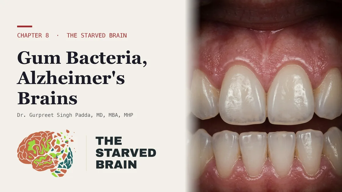
  • Dr. Gurpreet Singh Padda in a lab coat beside the title card reading The Gum Bacterium in 96% of Alzheimer's Brains, The Starved Brain, Chapter 8

    Gum Disease and Dementia: What Is Proven and What to Measure

    The Gum Bacterium in 96% of Alzheimer's Brains | The Starved Brain, Chapter 8

    Gum Disease and Dementia: What Is Proven and What to Measure

    Severe gum disease is tied to dementia; mild gum disease is not. Pooled studies found close to three times the odds of dementia with severe periodontitis and no measurable excess below moderate disease, and no one has shown that treating gum disease prevents dementia.

    A bacterium from bleeding gums turns up in most Alzheimer’s brains, and in plenty of healthy ones too. Reading that evidence correctly changes what you should worry about and what you should ask for.

    Is gum disease and dementia a real connection or a scary headline? The honest answer is both, depending on which gums and which claim. The chapter video, The Gum Bacterium in 96% of Alzheimer’s Brains, walks through the research on a mouth bacterium found in brain tissue. Before going further, one plain statement: Measura [Cardiometabolic and Autonomic Health Analysis] does not examine gums, does not test saliva, and has no test for any bacterium. That work belongs to a dentist or periodontist. What Measura can do is measure the person those gums belong to: blood sugar control, blood vessel function, and cognition.

    Is gum disease linked to dementia? The risk sits with severe disease

    Groups of researchers working independently kept finding the same thing: people with periodontal disease develop dementia more often. An umbrella review pooling that work put the extra risk at roughly a quarter and graded the certainty low to moderate. The detail that matters most is the gradient. Severe periodontitis came with close to three times the odds of dementia, while disease milder than moderate showed no measurable excess at all.

    So a little pink in the sink is a reason to book a dental visit. It is not a sign your memory is at risk. The exposure these studies point to is deep, bleeding, long-untreated infection, and it is common and quiet. A CDC analysis that examined every tooth in 10,683 American adults aged 30 to 79 found periodontitis in 42% and the severe form in 7.8%. Most people with it do not know.

    Is the gum bacterium found in Alzheimer’s brains?

    In 2019, a team looked for gingipains, the protein-cutting enzymes made by the gum bacterium Porphyromonas gingivalis, in brain samples. One of them turned up in 96% of Alzheimer’s samples, 51 of 53, and the amount tracked with markers of the disease. That is the headline. The same figure also shows the enzyme in 39% of samples from people without dementia, and the second gingipain in 52% of them. This organism is not a stranger to ordinary brains. What set Alzheimer’s tissue apart was how much was there.

    Two more facts belong beside that result. Twelve of the paper’s twenty-five authors worked for the company developing a gingipain-blocking drug, and no lab without a stake in that drug has repeated the brain staining. And a separate experiment in 20 ordinary mice, 10 exposed and 10 not, showed that painting the bacterium on the gums for 22 weeks put it in the brain alongside inflammation, amyloid and tau. That shows the path can run from mouth to brain in an animal. It measured tissue, not memory, and it has not been repeated.

    Does sugar feed the gum bacterium linked to Alzheimer’s?

    A popular claim says this bacterium survives only if you eat carbohydrates. Dr. Padda corrects it, even though it comes from people arguing the same side he does. P. gingivalis cannot use sugar for energy. It feeds on protein and pulls iron from blood. The real chain is less tidy and more convincing: a sugary diet feeds other mouth bacteria, shifts the balance, and inflames the gums. Inflamed, bleeding pockets deliver exactly the blood proteins this organism eats, and they open a door into the bloodstream.

    That door opens more often than people assume. Pooled studies found that toothbrushing puts bacteria into the blood in 8 to 26% of people, and plaque or gum inflammation roughly triples the odds. With healthy gums, that is trivial. With chronic periodontitis, it happens with an ordinary habit, day after day, for years. The gap is partly structural: teeth and the rest of the body are handled in separate offices, with separate charts, so the chronic wound in the mouth rarely appears in the medical record where cognition and blood sugar are tracked.

    Does treating gum disease prevent dementia?

    No one has shown that treating gum disease prevents dementia. The one large drug test, the GAIN trial, gave a gingipain blocker to 643 people with mild to moderate Alzheimer’s for 48 weeks and missed both main goals. Liver enzymes rose in 15% of the high-dose group against 2% on placebo, and the program was shut down in 2022. Among the 242 participants with the bacterium in their saliva, decline ran 57% slower on the higher dose, a subgroup result inside a failed trial, which is weak evidence. A follow-up trial will not report until 2029. Two genetic studies looking for a causal link found nothing that held up. And GAIN excluded people with poorly controlled diabetes or severe obesity, the metabolically complex patients most likely to be reading this.

    What Measura can measure in the same person

    The oral evidence cannot be collected at Measura. The body it happens in can be. A cognitive assessment sets a baseline for memory and thinking while you feel fine, so any later change has something to be compared against. Laboratory panels cover blood sugar control; in people with diabetes, a Cochrane review of 30 studies found that deep gum cleaning lowered HbA1c by about 0.43% at three to four months, so the dental chart and the A1C belong in the same conversation. And arterial stiffness and endothelial function testing looks at how stiff your arteries are and how their lining responds, the same territory a periodontal treatment trial tracked in people with severe gum disease. None of these tests detects gum disease. Together they show whether the chronic inflammation in your mouth is keeping company with trouble elsewhere.

    Questions for two appointments

    • At the dentist: ask for a full-mouth periodontal examination rather than a quick screening, and for your periodontal inflamed surface area, a figure in square millimeters calculated from probing depths and bleeding. Partial exams can miss more than half of disease.
    • At your physician: ask for your A1C to be read next to that dental chart, and ask about your 25-hydroxyvitamin D, which your immune cells use to make one of the body’s own antimicrobial peptides. No source sets a target level for that purpose.
    • On timing: do not have an inflammation marker such as hs-CRP drawn the day after a deep cleaning, because it rises briefly afterward.

    Treating the gums is worth doing on its own merits: you keep your teeth and lower a chronic inflammatory burden, whether or not the brain link is ever proven. Measurement tells you where you stand while that question waits. The complete evidence is in the book companion (the Chapter 8 companion page), and the earlier article in the series covers the sugar-driven inflammation behind this story (does sugar cause inflammation).

    Frequently asked questions

    Do bleeding gums mean I am going to get dementia?

    No. The pooled studies tie dementia risk to severe periodontitis, and gum disease milder than moderate showed no measurable excess. Bleeding when you brush is a reason to see a dentist soon, because it is treatable and it lets bacteria into the bloodstream more often. If memory is a worry, a cognitive baseline answers a different question than a dental exam does. More in memory and cognitive screening.

    Can Measura test me for P. gingivalis?

    No. Measura has no microbial, saliva or dental test. Saliva testing for the bacterium was done as part of a research drug trial and is not a routine clinical test. Your dentist or periodontist assesses the gums. Measura measures the metabolic, vascular and cognitive picture in the same person, which is useful alongside a dental chart but is not a replacement for one. A full list is in what Measura actually measures.

    Will treating my gum disease protect my memory?

    That has not been shown. The only large drug trial aimed at this bacterium failed, and genetic studies found no causal link that held up. Treatment is still worthwhile: it saves teeth, removes a chronic source of inflammation, and in people with diabetes it lowered A1C in pooled trials. The connection between the mouth and blood sugar is covered in insulin resistance and metabolic health.

    Why did I feel worse after a deep cleaning?

    That is the documented course. In a randomized trial of 120 people with severe periodontitis, intensive treatment briefly worsened blood vessel function and raised inflammatory markers at 24 hours, then improved vessel function by six months. Feeling off the next day is expected and passes. It is also why blood inflammation markers should not be drawn right after treatment. Vascular measurement is described on the arterial stiffness and endothelial function page.

    Why get a cognitive baseline if my memory is fine?

    Because change is only visible against a starting point. A score taken while you feel well turns a vague later worry into a comparison with real numbers. That matters for anyone with risks that build silently for decades, including chronic inflammation from the mouth or elsewhere. A baseline does not predict dementia; it makes future testing interpretable. More in what a cognitive baseline is for.

    Is there a link between brushing teeth and dementia?

    Brushing itself is not the problem. Pooled studies found that toothbrushing puts mouth bacteria into the blood in 8 to 26% of people, and plaque or gum inflammation roughly triples those odds. With healthy gums that is trivial. With chronic periodontitis it happens day after day for years, which is why the severe gum disease tied to dementia is the one worth treating.

    Can gum bacteria travel to the brain?

    In animals, yes. Painting the gum bacterium on the gums of mice for 22 weeks put it in the brain alongside inflammation, amyloid and tau. In people, one of its enzymes turned up in 96% of Alzheimer’s brain samples, and also in 39% of samples from people without dementia. What set Alzheimer’s tissue apart was how much was there, not whether it was there at all.

    Does sugar cause the gum disease linked to Alzheimer’s?

    Indirectly. P. gingivalis cannot use sugar for energy; it feeds on protein and pulls iron from blood. A sugary diet feeds other mouth bacteria, shifts the balance and inflames the gums. Those inflamed, bleeding pockets then supply the blood proteins this organism eats and open a door into the bloodstream. The inflammation side of that chain is covered in does sugar cause inflammation.

    Measure the body your gums belong to

    Request Measura testing for a cognitive baseline, blood sugar markers and vascular function, with results sent to your physician to read alongside your dental care.

    4477 Woodson Rd, Suite 201, St. Louis, MO 63134. Monday to Friday, 9:00 a.m. to 5:00 p.m. Please do not send symptoms, diagnoses or images through a web form — a website form is not a secure medical channel. Send your name and number and we will call you back.

    References

    • Qadir, B. H., Mahmood, M. K., Amin, Y. M. M., Kurda, H. A., Rasheed, T. A., Noori, A. H., Noori, Z. F., Abdulghafor, M. A., Fadhil, H. N. M., Fatih, M. T., Tardivo, D., Tassery, H., & Lan, R. (2025). Periodontitis and tooth loss are associated with higher risks of cognitive disorders: A systematic umbrella meta-analysis. Clinical and Experimental Dental Research, 11(6), Article e70240. https://doi.org/10.1002/cre2.70240
    • Kim, D.-H., & Han, G.-S. (2025). Periodontitis as a risk factor for dementia: A systematic review and meta-analysis. The Journal of Evidence-Based Dental Practice, 25(2), Article 102094. https://doi.org/10.1016/j.jebdp.2025.102094
    • Eke, P. I., Thornton-Evans, G. O., Wei, L., Borgnakke, W. S., Dye, B. A., & Genco, R. J. (2018). Periodontitis in US adults: National Health and Nutrition Examination Survey 2009–2014. Journal of the American Dental Association, 149(7), 576–588.e6. https://doi.org/10.1016/j.adaj.2018.04.023
    • Dominy, S. S., Lynch, C., Ermini, F., Benedyk, M., Marczyk, A., Konradi, A., Nguyen, M., Haditsch, U., Raha, D., Griffin, C., Holsinger, L. J., Arastu-Kapur, S., Kaba, S., Lee, A., Ryder, M. I., Potempa, B., Mydel, P., Hellvard, A., Adamowicz, K., … Potempa, J. (2019). Porphyromonas gingivalis in Alzheimer’s disease brains: Evidence for disease causation and treatment with small-molecule inhibitors. Science Advances, 5(1), Article eaau3333. https://doi.org/10.1126/sciadv.aau3333
    • Ilievski, V., Zuchowska, P. K., Green, S. J., Toth, P. T., Ragozzino, M. E., Le, K., Aljewari, H. W., O’Brien-Simpson, N. M., Reynolds, E. C., & Watanabe, K. (2018). Chronic oral application of a periodontal pathogen results in brain inflammation, neurodegeneration and amyloid beta production in wild type mice. PLoS ONE, 13(10), Article e0204941. https://doi.org/10.1371/journal.pone.0204941
    • Martins, C. C., Lockhart, P. B., Firmino, R. T., Kilmartin, C., Cahill, T. J., Dayer, M., Occhi-Alexandre, I. G. P., Lai, H., Ge, L., & Thornhill, M. H. (2023). Bacteremia following different oral procedures: Systematic review and meta-analysis. Oral Diseases, 30(3), 846–854. https://doi.org/10.1111/odi.14531
    • Tomás, I., Diz, P., Tobías, A., Scully, C., & Donos, N. (2011). Periodontal health status and bacteraemia from daily oral activities: Systematic review/meta-analysis. Journal of Clinical Periodontology, 39(3), 213–228. https://doi.org/10.1111/j.1600-051X.2011.01784.x
    • Simpson, T. C., Clarkson, J. E., Worthington, H. V., MacDonald, L., Weldon, J. C., Needleman, I., Iheozor-Ejiofor, Z., Wild, S. H., Qureshi, A., Walker, A., Patel, V. A., Boyers, D., & Twigg, J. (2022). Treatment of periodontitis for glycaemic control in people with diabetes mellitus. Cochrane Database of Systematic Reviews, 4(4), Article CD004714. https://doi.org/10.1002/14651858.CD004714.pub4
    • Tonetti, M. S., D’Aiuto, F., Nibali, L., Donald, A., Storry, C., Parkar, M., Suvan, J., Hingorani, A. D., Vallance, P., & Deanfield, J. (2007). Treatment of periodontitis and endothelial function. New England Journal of Medicine, 356(9), 911–920. https://doi.org/10.1056/NEJMoa063186
    • Nesse, W., Abbas, F., van der Ploeg, I., Spijkervet, F. K. L., Dijkstra, P. U., & Vissink, A. (2008). Periodontal inflamed surface area: Quantifying inflammatory burden. Journal of Clinical Periodontology, 35(8), 668–673. https://doi.org/10.1111/j.1600-051X.2008.01249.x

    Related reading

  • Dr. Gurpreet Singh Padda beside the Chapter 5 title card of The Starved Brain reading Your Lab Says Normal. Your Brain Is Shrinking.

    Methylmalonic Acid Test: When a Normal B12 Is Not Enough

    Your Lab Says Normal. Your Brain Is Shrinking. | The Starved Brain, Chapter 5

    Methylmalonic Acid Test: When a Normal B12 Is Not Enough

    A methylmalonic acid test shows whether B12 is doing its job inside your cells, not just how much is floating in your blood. When B12 fails at that job, methylmalonic acid backs up, so the test can flag a shortfall behind a normal serum B12.

    Your B12 came back normal. In an Oxford brain-imaging study, so did the results of the people losing brain volume fastest, and the methylmalonic acid test is one way to see what that number leaves out.

    The video above, Your Lab Says Normal. Your Brain Is Shrinking., carries a warning most people never hear at a physical: a serum B12 inside the reference range does not prove your cells are getting enough. The methylmalonic acid test asks a different question. Instead of counting how much B12 is floating in your blood, it looks for a chemical backlog that builds up when B12 is failing at its job inside the cell.

    Dr. Padda spent years telling people with prediabetes that an A1C that was “not too bad” was fine. He was wrong, and a B12 in the normal range invites exactly the same shrug. It deserves the same second look.

    Can you be low in B12 with a normal B12 level?

    A laboratory range is drawn by testing a big group of people presumed healthy and labeling the middle of their results as normal. Nobody checks whether that group was well nourished first. B12 is made by bacteria and comes to people almost entirely from animal foods, so a population that eats fewer of them pulls its own range down, and the report keeps printing the word normal. The range tells you where the crowd stands. It does not tell you where health begins.

    Researchers at Oxford put that to the test in 107 cognitively normal adults aged 61 to 87, scanning their brains every year for five years. Not one was deficient by the usual laboratory cutoff. Even so, people in the bottom third of B12, below 308 pmol/L, had about six times the odds of landing in the group losing brain volume fastest. A larger cohort of 501 older adults in Stockholm later pointed the same way: higher B12 went with slower shrinkage, and higher homocysteine went with faster.

    What does a methylmalonic acid test measure?

    Only two enzymes in the human body use B12. When one of them stalls, methylmalonic acid backs up. When the other stalls, homocysteine backs up. That is why both are called functional markers. Serum B12 counts the inventory on the shelf; methylmalonic acid and homocysteine tell you whether the work got done. A third blood test, holotranscobalamin, measures the small active share of B12 that cells can take up. About 80% of the B12 a standard test counts is bound to a carrier protein that does not hand it to your tissues.

    The gap between the shelf and the work is measurable. Among 548 surviving members of the original Framingham cohort, methylmalonic acid was markedly raised in 11.3%, and the authors concluded that many older people with normal serum levels were metabolically deficient. In a laboratory study of people with cognitive complaints, total serum B12 did little better than a coin toss at picking out deficiency, while holotranscobalamin did better, though still modestly.

    Who is most likely to have a B12 shortfall with a normal level?

    • Adults over 60. Freeing B12 from food takes stomach acid and intrinsic factor, and in older people trouble releasing B12 from food becomes the leading cause of deficiency.
    • People taking metformin. In the Diabetes Prevention Program, low or borderline B12 reached 19.1% in the metformin group against 9.5% on placebo at five years.
    • People on long-term acid suppression. In a large Kaiser Permanente study, two or more years of a proton pump inhibitor carried higher odds of a B12 deficiency diagnosis, though studies that pooled the data call the average effect small.
    • People who eat few animal foods, whose intake starts low.

    None of these is a reason to stop a medication. Each is a reason to know the number, and to know what the number leaves out.

    Why can one B12 marker read alone mislead you?

    VITACOG randomized 271 adults aged 70 and over with mild cognitive impairment to high-dose B vitamins or placebo. Over two years, whole-brain shrinkage on MRI ran 29.6% slower on the vitamins, and the benefit concentrated in people whose homocysteine was high at the start. Then the investigators sorted participants by blood omega-3 levels. In the top third, the vitamins slowed atrophy. In the bottom third, they had no significant effect.

    That split was an analysis done after the trial, so it is a hypothesis with a mechanism, not a settled law. The mechanism is easy to picture. B vitamins run the assembly line that makes phosphatidylcholine for cell membranes and myelin, and omega-3 fats are among the parts that line installs. Run the line with no parts and nothing ships. The practice position is that the two are measured and treated as a pair. For your lab work, the lesson is sharper than it looks: a partner nutrient can decide whether the first one matters at all, so one marker read alone can steer you wrong.

    The food supply produces both shortfalls at the same time. It delivers abundant calories while crowding out the animal foods that carry B12 and the fish that carries EPA and DHA, then floods the plate with competing omega-6 fats. A person can be overfed and underbuilt in one body, and neither shows on the scale. The pattern also runs into insulin resistance, because the drug most often handed to insulin-resistant patients is the one that lowers B12.

    Where Measura fits, and where it does not

    Measura [Cardiometabolic and Autonomic Health Analysis] exists to put numbers on what a routine visit skips. It takes measurements, sends every finding to your physician, and leaves diagnosis and treatment decisions with that physician. Blood markers are drawn through laboratory panels, and which markers go on the order is a decision you make with your physician. Asking by name for methylmalonic acid, homocysteine and holotranscobalamin next to serum B12 is a reasonable way to open that conversation.

    A shortfall that has run quietly for years does not always announce itself, so two other measurements show what the rest of the system is doing. A cognitive assessment records a baseline for memory and thinking before anyone is in a position to call a change a diagnosis. Sudomotor testing measures sweat-gland function in the small nerve fibers of the hands and feet, and nerve health belongs in the same conversation: in the Diabetes Prevention Program, neuropathy was more common in metformin users whose B12 was low. Neither test measures B12. Each shows the terrain while the blood work answers the nutrient question.

    What should you ask your doctor about B12 testing?

    • Where does my B12 sit inside the range, not just whether it is flagged?
    • Can we add methylmalonic acid and homocysteine, and holotranscobalamin if the laboratory offers it?
    • Can my folate be read beside my B12? Folate can correct the blood count while a B12 shortfall keeps affecting nerves, which is why it helps to know B12 status before taking high-dose folate.
    • What is my omega-3 index?
    • Am I taking metformin or an acid suppressor, and when was my B12 last checked?

    More ideas for that visit are on questions worth asking your doctor. The book companion for Chapter 5 of The Starved Brain lists every study and number behind these questions, and the next post in the series, on B12 in aging parents, follows the same shortfall into later life. Unmeasured is unmanaged, and a normal result should start a question rather than end one.

    Frequently asked questions

    What does a high methylmalonic acid result mean?

    A raised methylmalonic acid level suggests B12 is not getting its work done inside your cells, even when the serum B12 number looks fine. It is one piece of evidence, not a diagnosis by itself. Your physician reads it with serum B12, holotranscobalamin, homocysteine, your medications and your history. For how a result fits into the full picture, see understanding your results.

    Is a homocysteine test the same as a methylmalonic acid test?

    No. Both rise when the B12 machinery stalls, but homocysteine also rises when folate runs short, so the two answer slightly different questions. Homocysteine carries its own weight: among 1,092 Framingham adults with a mean age of 76, levels above 14 µmol/L went with nearly double the Alzheimer’s risk. For what any single marker can and cannot show, read what a test result can tell you.

    Can a normal B12 level still affect memory?

    In the Oxford cohort, lower B12 inside the normal range was linked to faster brain shrinkage in people with no cognitive impairment. That is an association, not proof that B12 caused it, and the study was small. It is a strong reason to look at where your number sits and to record a memory baseline early. See memory and cognitive screening.

    Does Measura run B12 blood work?

    Blood markers at Measura are drawn through laboratory panels, and the markers on the order are chosen with your physician. Measura measures and sends findings to your physician; it does not treat deficiency or prescribe supplements. If you want to know which outside laboratory processes the samples, read which laboratory runs the blood work.

    How often should B12 be checked if I take metformin?

    No fixed schedule appears in the research summarized here, but the risk builds with time: in the Diabetes Prevention Program, each added year of metformin carried an odds ratio of 1.13 for low B12. That makes a repeat measurement a conversation worth having at regular visits. For how repeat testing is usually planned, see how often to repeat testing.

    Is methylmalonic acid high or low in B12 deficiency?

    High. When B12 fails at its job inside the cell, one of the two enzymes that depend on it stalls and methylmalonic acid backs up. That is why the test can flag a shortfall while serum B12 still reads normal. Among 548 surviving members of the original Framingham cohort, methylmalonic acid was markedly raised in 11.3%, and many older people with normal serum levels were metabolically deficient.

    How are vitamin B12 and methylmalonic acid related?

    Only two enzymes in the human body use B12. When one stalls, methylmalonic acid builds up; when the other stalls, homocysteine builds up. Serum B12 counts the inventory on the shelf, while methylmalonic acid shows whether the work got done. Holotranscobalamin adds a third view: the small active share of B12 that cells can actually take up.

    What causes a B12 shortfall that raises methylmalonic acid?

    The common routes are age, medication and diet. After 60, trouble releasing B12 from food becomes the leading cause of deficiency. In the Diabetes Prevention Program, low or borderline B12 reached 19.1% on metformin against 9.5% on placebo at five years. Long-term acid suppression and eating few animal foods also lower it. None of these is a reason to stop a medication.

    Ask for more than one number

    Request Measura testing and bring your B12 questions with you, so your results reach your physician with the context they need.

    4477 Woodson Rd, Suite 201, St. Louis, MO 63134. Monday to Friday, 9:00 a.m. to 5:00 p.m. Please do not send symptoms, diagnoses or images through a web form — a website form is not a secure medical channel. Send your name and number and we will call you back.

    References

    • Vogiatzoglou, A., Refsum, H., Johnston, C., Smith, S. M., Bradley, K. M., de Jager, C., Budge, M. M., & Smith, A. D. (2008). Vitamin B12 status and rate of brain volume loss in community-dwelling elderly. Neurology, 71(11), 826–832. https://doi.org/10.1212/01.wnl.0000325581.26991.f2
    • Hooshmand, B., Mangialasche, F., Kalpouzos, G., Solomon, A., Kåreholt, I., Smith, A. D., Refsum, H., Wang, R., Mühlmann, M., Ertl-Wagner, B., Laukka, E. J., Bäckman, L., Fratiglioni, L., & Kivipelto, M. (2016). Association of vitamin B12, folate, and sulfur amino acids with brain magnetic resonance imaging measures in older adults: A longitudinal population-based study. JAMA Psychiatry, 73(6), 606–613. https://doi.org/10.1001/jamapsychiatry.2016.0274
    • Herrmann, W., & Obeid, R. (2012). Cobalamin deficiency. Sub-Cellular Biochemistry, 56, 301–322. https://doi.org/10.1007/978-94-007-2199-9_16
    • Lindenbaum, J., Rosenberg, I. H., Wilson, P. W., Stabler, S. P., & Allen, R. H. (1994). Prevalence of cobalamin deficiency in the Framingham elderly population. The American Journal of Clinical Nutrition, 60(1), 2–11. https://doi.org/10.1093/ajcn/60.1.2
    • Murphy, M. J., Brandie, F., Ebare, M., Harrison, M., Dow, E., Bartlett, W. A., & Craig, D. (2021). Personalising laboratory medicine in the ‘real world’: Assessing clinical utility, by clinical indication, of serum total B12 and Active-B12 (holotranscobalamin) in the diagnosis of vitamin B12 deficiency. Annals of Clinical Biochemistry, 58(5), 445–451. https://doi.org/10.1177/00045632211003605
    • Aroda, V. R., Edelstein, S. L., Goldberg, R. B., Knowler, W. C., Marcovina, S. M., Orchard, T. J., Bray, G. A., Schade, D. S., Temprosa, M. G., White, N. H., & Crandall, J. P., for the Diabetes Prevention Program Research Group (2016). Long-term metformin use and vitamin B12 deficiency in the Diabetes Prevention Program Outcomes Study. The Journal of Clinical Endocrinology & Metabolism, 101(4), 1754–1761. https://doi.org/10.1210/jc.2015-3754
    • Smith, A. D., Smith, S. M., de Jager, C. A., Whitbread, P., Johnston, C., Agacinski, G., Oulhaj, A., Bradley, K. M., Jacoby, R., & Refsum, H. (2010). Homocysteine-lowering by B vitamins slows the rate of accelerated brain atrophy in mild cognitive impairment: A randomized controlled trial. PLoS ONE, 5(9), e12244. https://doi.org/10.1371/journal.pone.0012244
    • Jernerén, F., Elshorbagy, A. K., Oulhaj, A., Smith, S. M., Refsum, H., & Smith, A. D. (2015). Brain atrophy in cognitively impaired elderly: The importance of long-chain ω-3 fatty acids and B vitamin status in a randomized controlled trial. The American Journal of Clinical Nutrition, 102(1), 215–221. https://doi.org/10.3945/ajcn.114.103283
    • Seshadri, S., Beiser, A., Selhub, J., Jacques, P. F., Rosenberg, I. H., D’Agostino, R. B., Wilson, P. W. F., & Wolf, P. A. (2002). Plasma homocysteine as a risk factor for dementia and Alzheimer’s disease. The New England Journal of Medicine, 346(7), 476–483. https://doi.org/10.1056/NEJMoa011613
    • Lam, J. R., Schneider, J. L., Zhao, W., & Corley, D. A. (2013). Proton pump inhibitor and histamine 2 receptor antagonist use and vitamin B12 deficiency. JAMA, 310(22), 2435–2442. https://doi.org/10.1001/jama.2013.280490

    Related reading

  • Title card for The Starved Brain Chapter 4 video: Dr. Gurpreet Singh Padda with the title Your Brain Is Built From the Fat You Were Told to Fear

    Statins and Memory Loss: What to Measure Before Anyone Guesses

    Your Brain Is Built From the Fat You Were Told to Fear | The Starved Brain, Chapter 4

    Statins and Memory Loss: What to Measure Before Anyone Guesses

    Whether a statin is affecting your memory can be measured rather than guessed. Large trials found no cognitive harm on average, the FDA label describes rare, reversible memory complaints, and a repeatable cognitive test turns the worry into a measurement.

    Your lipid panel reports on the cholesterol in your blood. The cholesterol your brain is built from is made on site, behind a barrier, and no routine blood test reads it.

    Worries about statins and memory loss usually arrive with nothing attached: a name that will not come, a word lost mid-sentence, a sense of being slower since the prescription started. Chapter 4 of The Starved Brain by Dr. Gurpreet Singh Padda, MD, MBA, MHP, and the video that goes with it, take that worry seriously without turning it into an instruction. Nothing here is a reason to stop a prescribed medication on your own. The point for Measura [Cardiometabolic and Autonomic Health Analysis] is different: a worry cannot be tested, and a measurement can.

    Where does the brain get its cholesterol?

    Your brain is about 2% of your body weight and holds roughly a quarter of the body’s cholesterol. It uses that cholesterol as building material. Myelin, the insulation around nerve fibers that lets signals travel fast, is roughly 70–80% fat by dry weight, and the supply of cholesterol sets how quickly it can be made. Synapses, the connections where memories live, need cholesterol to form.

    Here is the part most patients never hear. Your brain does not take its cholesterol from your blood. The LDL and HDL particles on a cholesterol report cannot cross the blood–brain barrier. Brain cholesterol is made inside the brain, mostly by support cells called astrocytes, and it is replaced slowly. So your lipid panel, however carefully read, says very little about the cholesterol your neurons depend on. It measures a different compartment.

    Do statins affect memory, and what does the evidence show?

    Statins lower cholesterol by blocking an enzyme called HMG-CoA reductase. Every cell uses that enzyme, including astrocytes. Fat-soluble statins such as simvastatin, atorvastatin and lovastatin enter the brain more easily than water-soluble ones such as pravastatin and rosuvastatin. That is real pharmacology. It has not shown up as a penalty in large population studies: when researchers sorted statins by solubility, the fat-soluble drugs did no worse for dementia.

    In 2012 the FDA added wording to every statin label about rare reports of memory loss, forgetfulness and confusion, generally not serious and reversible once the drug was stopped. The label describes symptoms starting anywhere from a day to years after the first dose and clearing at a median of three weeks after stopping. It records a pattern in reports, not how often it happens. Randomized trials involving well over a hundred thousand people found no cognitive harm on average.

    Then there is the small study that makes the question testable. In 2012, researchers took 18 people with Alzheimer’s disease who were on statins, measured their thinking, stopped the statin for six weeks, and restarted it for six weeks. A 30-point memory screening test improved by about 2 points off the drug and dropped by about the same amount back on it. A longer set of thinking tests did not change. It was a pilot with no control group, and a 2-point shift sits inside the normal variation of that screening test. It is a signal, not proof, and it points to a possible effect in a susceptible few rather than in most people taking a statin.

    Dr. Padda spent years as a strict vegetarian defending the dietary advice that told people to fear eggs and animal fat, and he abandoned that position when the physiology did not hold up. That history is why the argument here rests on measurement rather than conviction: the answer for one person comes from testing that person.

    What does a routine visit miss about statins and memory?

    When a patient on a statin mentions memory, a routine visit usually has two tools: a lipid panel and a conversation. The lipid panel does not see brain cholesterol. The conversation depends on how well you remember what you used to remember, which is the very thing in question. Add a short appointment, a long medication list, and a system that files memory complaints under getting older, and the most common result is a shrug.

    What is missing is a baseline. Without a record of how you performed before a change, nobody can say whether you have changed.

    How can memory changes on a statin be measured?

    Measura measures; it does not diagnose disease on its own or manage medication, and results go to your physician. For this question, the useful measurements are:

    • Cognitive assessment: a structured record of memory, attention and thinking speed. Repeated the same way, it lets you and your physician compare you with yourself over time, including before and after any change your physician makes to your treatment.
    • Laboratory panels: blood work describing metabolic health beyond cholesterol. This matters because in blinded statin trials, about 62% of new diabetes cases occurred in people whose blood sugar was already in the top quarter at the start.
    • Arterial stiffness and endothelial function: the vascular side, which is why most people take a statin in the first place. It speaks to the heart question rather than the brain one, and keeping the two apart makes both conversations clearer.
    • Bioimpedance body composition: muscle and fat measured separately, part of the metabolic picture a scale cannot give.

    What should you ask your doctor about statins and memory?

    Memory changes that began on a statin deserve investigation rather than assumption, and the investigation belongs to you and the physician who prescribed it. Useful questions:

    • Did my memory or thinking change after I started this medication, and can we document it?
    • Can we record a cognitive baseline now, with a test we can repeat the same way later?
    • My cholesterol numbers look good. What do we know about my blood sugar and insulin?
    • If we ever check whether the medication is involved, how would we do it safely, and who decides?

    Getting ready for testing is covered in how to prepare. The full evidence, including the trials on both sides, is in the book companion for Chapter 4, and the fuel side of the same book is in type 3 diabetes and the normal blood sugar test.

    The larger point about building materials

    The brain is built from lipids: cholesterol, saturated fat, phospholipids and the long-chain fats DHA and EPA. For fifty years, public health advice pushed people away from the foods richest in them. In a Finnish cohort followed for about twenty-two years, more dietary cholesterol was not linked to more dementia, and in the Rush Memory and Aging Project, people who ate more than one egg a week had about half the risk of Alzheimer’s dementia. Both are observational. Neither proves that eggs protect the brain, and both run opposite to the warning most people grew up with. Unmeasured is unmanaged, and the brain’s construction supply is one of the least measured systems in routine care.

    Frequently asked questions

    Can statins cause memory loss?

    The FDA label for every statin describes rare reports of memory loss and confusion that were generally not serious and went away after stopping. Large randomized trials found no cognitive harm on average. A small pilot suggested some people with existing cognitive impairment may be affected, which is why individual testing under a physician’s supervision is the useful question. See memory and cognitive screening.

    Does my cholesterol test show my brain’s cholesterol?

    No. Cholesterol particles in the blood do not cross the blood–brain barrier, and the brain makes nearly all of its own supply. A lipid panel describes your blood and your cardiovascular picture. It says very little about the cholesterol used to build and maintain myelin and synapses. How results are read is covered in understanding your results.

    Should I stop my statin if I notice memory changes?

    Not on your own. Statins reduce heart attacks and strokes in appropriately selected patients, and stopping without supervision can cause real harm. Bring the change to the physician who prescribed the medication, ideally with a documented cognitive baseline, so any decision rests on measurement rather than memory. Help framing that visit is in questions worth asking your doctor.

    What does a cognitive assessment actually measure?

    It uses structured tasks to record memory, attention, language and thinking speed, scored the same way each time. It does not diagnose Alzheimer’s disease or any other condition on its own. Its value is comparison: a later result can be set against your own earlier one. Details are on the cognitive assessment page.

    Why does blood sugar matter for someone taking a statin?

    In blinded statin trials, extra cases of new diabetes clustered in people whose blood sugar was already near the top of the normal range. Knowing whether insulin resistance is present gives you and your physician a fuller picture of the tradeoffs involved in your care. More is in insulin resistance and metabolic health.

    Which statin is least likely to cause memory loss?

    Fat-soluble statins such as simvastatin, atorvastatin and lovastatin enter the brain more easily than water-soluble ones such as pravastatin and rosuvastatin. That is real pharmacology, yet it has not shown up as a penalty in large population studies: sorted by solubility, the fat-soluble drugs did no worse for dementia. The answer for one person comes from measuring that person, starting with a cognitive baseline.

    Is memory loss from statins reversible?

    On the FDA label, yes. Since 2012 every statin label describes rare reports of memory loss, forgetfulness and confusion that were generally not serious and cleared once the drug was stopped, at a median of three weeks. Any change to the prescription belongs to the physician who wrote it, and a documented cognitive baseline lets the two of you see whether anything actually changed.

    How soon after starting a statin can memory problems begin?

    The FDA label describes symptoms starting anywhere from a day to years after the first dose. That wide window is why recall alone cannot settle the question. A structured cognitive assessment recorded now, and repeated the same way later, compares you with yourself instead of with what you remember about how sharp you used to be.

    Turn a memory worry into a measurement

    Request Measura testing to record a cognitive and metabolic baseline, with the results sent to the physician who manages your care.

    4477 Woodson Rd, Suite 201, St. Louis, MO 63134. Monday to Friday, 9:00 a.m. to 5:00 p.m. Please do not send symptoms, diagnoses or images through a web form — a website form is not a secure medical channel. Send your name and number and we will call you back.

    References

    • Dietschy, J. M., & Turley, S. D. (2004). Thematic review series: Brain lipids. Cholesterol metabolism in the central nervous system during early development and in the mature animal. Journal of Lipid Research, 45(8), 1375–1397. https://doi.org/10.1194/jlr.R400004-JLR200
    • Saher, G., Brügger, B., Lappe-Siefke, C., Möbius, W., Tozawa, R., Wehr, M. C., Wieland, F., Ishibashi, S., & Nave, K.-A. (2005). High cholesterol level is essential for myelin membrane growth. Nature Neuroscience, 8(4), 468–475. https://doi.org/10.1038/nn1426
    • Padala, K. P., Padala, P. R., McNeilly, D. P., Geske, J. A., Sullivan, D. H., & Potter, J. F. (2012). The effect of HMG-CoA reductase inhibitors on cognition in patients with Alzheimer’s dementia: A prospective withdrawal and rechallenge pilot study. The American Journal of Geriatric Pharmacotherapy, 10(5), 296–302. https://doi.org/10.1016/j.amjopharm.2012.08.002
    • U.S. Food and Drug Administration / Bristol-Myers Squibb. (2012). PRAVACHOL (pravastatin sodium) tablets: Prescribing information, Section 6.2 Postmarketing Experience (label revised with the statin-class cognitive wording; FDA Reference ID 3090896). https://www.accessdata.fda.gov/drugsatfda_docs/label/2012/019898s062lbl.pdf
    • Richardson, K., Schoen, M., French, B., Umscheid, C. A., Mitchell, M. D., Arnold, S. E., Heidenreich, P. A., Rader, D. J., & deGoma, E. M. (2013). Statins and cognitive function: A systematic review. Annals of Internal Medicine, 159(10), 688–697. https://doi.org/10.7326/0003-4819-159-10-201311190-00007
    • Olmastroni, E., Molari, G., De Beni, N., Colpani, O., Galimberti, F., Gazzotti, M., Zambon, A., Catapano, A. L., & Casula, M. (2022). Statin use and risk of dementia or Alzheimer’s disease: A systematic review and meta-analysis of observational studies. European Journal of Preventive Cardiology, 29(5), 804–814. https://doi.org/10.1093/eurjpc/zwab208
    • Cholesterol Treatment Trialists’ (CTT) Collaboration. (2024). Effects of statin therapy on diagnoses of new-onset diabetes and worsening glycaemia in large-scale randomised blinded statin trials: An individual participant data meta-analysis. The Lancet Diabetes & Endocrinology, 12(5), 306–319. https://doi.org/10.1016/S2213-8587(24)00040-8
    • Cholesterol Treatment Trialists’ Collaboration. (2026). Assessment of adverse effects attributed to statin therapy in product labels: A meta-analysis of double-blind randomised controlled trials. The Lancet, 407(10529), 689–703. https://doi.org/10.1016/S0140-6736(25)01578-8
    • Ylilauri, M. P. T., Voutilainen, S., Lönnroos, E., Mursu, J., Virtanen, H. E. K., Koskinen, T. T., Salonen, J. T., Tuomainen, T.-P., & Virtanen, J. K. (2017). Association of dietary cholesterol and egg intakes with the risk of incident dementia or Alzheimer disease: The Kuopio Ischaemic Heart Disease Risk Factor Study. American Journal of Clinical Nutrition, 105(2), 476–484. https://doi.org/10.3945/ajcn.116.146753
    • Pan, Y., Wallace, T. C., Karosas, T., Bennett, D. A., Agarwal, P., & Chung, M. (2024). Association of egg intake with Alzheimer’s dementia risk in older adults: The Rush Memory and Aging Project. The Journal of Nutrition, 154(7), 2236–2243. https://doi.org/10.1016/j.tjnut.2024.05.012

    Related reading

  • Video thumbnail for "Which Laboratory Performs the Testing?" with Dr. Gurpreet Singh Padda, MD, MBA, MHP

    Which Laboratory Runs the Blood Work

    Which Laboratory Performs the Testing?

    How the laboratory work is handled

    Most of the Measura suite is performed in the office with non-invasive equipment. Where blood work is part of the plan, it is drawn at the same visit and processed by a clinical laboratory.

    Which blood tests are ordered, and why not a fixed panel?

    Panels are selected to the clinical question rather than ordered as a standard bundle. Someone being assessed for burning feet needs glycemic markers and vitamin B12; someone being assessed for a stalled nutrition plan needs insulin, glucose and a lipid particle analysis. Ordering everything on the menu produces incidental findings that generate work and answer nothing.

    The laboratory panels page lists the available groups.

    What does a standard annual blood panel leave out?

    • Fasting insulin, alongside glucose and hemoglobin A1c. Insulin rises for years before glucose does, so measuring only glucose describes a late stage of the process.
    • C-peptide and fructosamine, which characterize the same process differently.
    • Inflammatory markers including high-sensitivity C-reactive protein, ferritin and homocysteine.
    • Lipid particle analysis and oxidized LDL rather than total cholesterol alone.
    • A full thyroid panel — free T3, T4, reverse T3 and TSH, rather than TSH in isolation.
    • Vitamin B12, red cell magnesium, zinc and the omega ratio. B12 deficiency alone can produce both neuropathy and cognitive change, and it is correctable.

    Do you need to fast before blood work?

    Glucose, insulin, triglycerides and every index derived from them are shifted by a recent meal. The fasting state is documented rather than assumed, because a non-fasting sample reported as fasting is worse than no sample. Water is allowed and encouraged.

    Draw time is recorded too. Cortisol, testosterone and thyroid markers all vary through the day, so a comparison between two draws at different times is not really a comparison.

    Can I bring my own recent lab results?

    Bring them. Recent outside laboratory work is reconciled rather than duplicated wherever it answers the same question. That is one of the more common ways to shorten a visit.

    How are the blood test results interpreted?

    Against the in-office measurements, not separately. An abnormal sudomotor result with a low B12 points somewhere quite specific; the same result with a raised hemoglobin A1c points somewhere else. Neither on its own tells you which. How to read your report.

    Frequently asked questions

    Where is the blood work done?

    Most of the Measura suite is performed in the office with non-invasive equipment. When blood work is part of your plan, it is drawn at the same visit, so there is no separate trip, and the sample is processed by a clinical laboratory. Recent outside results are reconciled rather than repeated wherever they answer the same question.

    Can I drink water before fasting blood work?

    Yes. Water is allowed and encouraged. What matters is food, because glucose, insulin, triglycerides and every index derived from them are shifted by a recent meal. The fasting state is written down rather than assumed, since a non-fasting sample reported as fasting is worse than no sample at all.

    Does the time of day matter for a blood test?

    Yes, for some markers. Cortisol, testosterone and thyroid markers all change through the day, so the draw time is recorded with every sample. Comparing two draws taken at different times of day is not really a comparison, which is why the time is kept alongside the result.

    Related reading

  • Video thumbnail for "How Measura Gathers Actionable Insights" with Dr. Gurpreet Singh Padda, MD, MBA, MHP

    How to Read Your Measura Report

    How Measura Gathers Actionable Insights

    How to read your report

    Three habits make a report useful instead of alarming: read the pattern, read the conditions, and keep the previous one.

    The report is organized by body system

    Not by machine. Circulation, small nerve fibers, autonomic function, body composition, metabolism, balance and cognition each get a section, and each section answers a different question. Understanding your results sets out what each one asks.

    Habit one: read across sections, not down one

    The most useful information is almost always in a combination. Reduced conductance in the feet plus a reduced toe index plus dry, cracked skin is a specific picture with a specific action attached. Any one of those three on its own is much less informative.

    The same is true in the other direction. A normal ankle-brachial index next to a damped waveform is not a contradiction — it usually means the arteries are too calcified to compress, and the waveform is the one telling the truth.

    Habit two: read the conditions along with the numbers

    Every page should carry the conditions the measurements were taken under: whether you fasted, whether you had caffeine or nicotine, how you slept, whether you had alcohol the night before, the time of day, and your medication list. Those are not footnotes. Several of them move the results more than mild disease would.

    Habit three: keep it, and bring it next time

    The single most valuable output of this suite is the comparison between one assessment and the next. That only works if somebody kept the first one. Ask for a copy every time and keep them together.

    What should I do if a result is abnormal?

    1. Do not act on it alone. Read it against the rest of the report and against the conditions.
    2. If it is borderline, expect it to be repeated under controlled conditions before it changes anything.
    3. If it is clearly abnormal and consistent with the rest of the picture, that is the conversation to have with a clinician who knows your history.
    4. Never stop or change a medication because a number looked wrong.

    What should I do with a normal report?

    Keep it. A complete normal baseline in a person in the risk group is exactly what makes a future measurement interpretable, and it is the most common outcome for someone who comes in early rather than late.

    Frequently asked questions

    How should I interpret my test results?

    Use three habits. Read across sections rather than down one, because the most useful information is usually a combination of findings. Read the testing conditions along with the numbers. And keep your previous report, because comparing one assessment with the next is the most valuable thing the testing produces. Then go over it with a clinician who knows your history.

    Can caffeine, food or sleep change my test results?

    Yes. Whether you fasted, had caffeine or nicotine, slept poorly or drank alcohol the night before, plus the time of day and your medications, can all move the results. Several of them move the numbers more than mild disease would. That is why each page of the report should record the conditions the measurements were taken under.

    Should I keep my old test reports?

    Yes. The single most valuable output of the testing is the comparison between one assessment and the next, and that only works if somebody kept the first report. Ask for a copy every time, keep them together and bring them with you to your next assessment so the new numbers can be read against the old ones.

    Related reading

  • Video thumbnail for "How Often Should Measura Testing Be Repeated?" with Dr. Gurpreet Singh Padda, MD, MBA, MHP

    How Often Should You Repeat Cardiometabolic Testing?

    How Often Should Measura Testing Be Repeated?

    How often should testing be repeated?

    Repeat cardiometabolic testing about once a year when a study is normal and nothing is being changed, within weeks when a result is borderline, and every three to six months for metabolic measures when an abnormal result is being actively treated.

    Set the interval from what was found and what is being changed, not from the calendar.

    The four situations

    SituationTypical intervalWhy
    Normal study, risk group, nothing being changedAbout a yearThe value is the trend; annual keeps it comparable without over-testing
    Borderline resultWeeks, under controlled conditionsBorderline values are frequently artifacts of preparation, sleep or coaching. Repeat before concluding
    Abnormal result, something being actively changedThree to six months for metabolic measuresLong enough for a real change, short enough to correct course
    Abnormal structural vascular findingLonger, and guided by the clinical planStructural change is slower than metabolic change
    Intervals are set clinically. These are the shapes, not rules.

    Why not retest on a fixed calendar schedule?

    The point of repeating is comparison, and comparison only works when something has had a chance to move. Re-measuring resting energy expenditure six weeks into a nutrition plan tells you almost nothing; re-measuring at four months tells you whether the plan is working. Conversely, waiting a full year to recheck a borderline ankle-brachial index in a smoker with worsening claudication is too long.

    How do you make a repeat test comparable?

    A repeat measured under different conditions is not a comparison. Same fasting state, same time of day, no caffeine or nicotine either time, same instrument, same technique, same posture and the same rest period. If the first was taken after a poor night’s sleep, note it and take that into account rather than pretending the two are equivalent.

    This is why the conditions are recorded as part of the result. Why acquisition conditions matter.

    Which results change fastest after treatment?

    • Within weeks to months: triglycerides, fasting glucose and insulin, measured metabolic rate, body composition, heart rate variability.
    • Within months to a year: waist circumference, hemoglobin A1c, inflammatory markers, endothelial markers, sudomotor conductance where a driver has been corrected.
    • Slowly, if at all: established arterial stiffness and structural arterial narrowing.

    That ordering is also the argument for measuring early: the things that respond fastest are the ones upstream of the things that barely respond at all.

    Frequently asked questions

    Is it normal to repeat testing every year?

    Yes, when the first study was normal, you are in a risk group and nothing is being changed. Repeating about once a year keeps the results comparable without over-testing, because the value is in the trend. A borderline or abnormal result changes that schedule: borderline results are repeated within weeks, and abnormal results under active treatment are rechecked sooner.

    How long should you wait between tests?

    It depends on what was found and what is being changed. A borderline result is repeated within weeks under controlled conditions. An abnormal result that is being treated is rechecked every three to six months for metabolic measures. An abnormal structural finding in the arteries is rechecked on a longer schedule set by the clinical plan, because structure changes slowly.

    What are common cardiometabolic biomarkers?

    The measures covered here include triglycerides, fasting glucose and insulin, hemoglobin A1c, inflammatory and endothelial markers, measured metabolic rate, body composition, waist circumference, heart rate variability, sudomotor conductance and arterial stiffness. They change at different speeds: triglycerides, glucose and metabolic rate can move within weeks to months, while established arterial stiffness changes slowly, if at all.

    How soon after starting a new plan should you retest?

    Give the plan time to work before measuring again. Rechecking resting energy expenditure six weeks into a nutrition plan tells you almost nothing, while rechecking at four months shows whether the plan is working. For metabolic measures under active treatment, three to six months is long enough for a real change and short enough to correct course.

    Related reading