Category: Nerves and neuropathy

  • Dr. Gurpreet Singh Padda presenting the title card Nerve or Joint? Pain Speaks Two Languages, The Pained Brain, Chapter 20

    Nerve Pain After Knee Replacement: What Testing Can and Cannot Show

    Nerve or Joint? Pain Speaks Two Languages | The Pained Brain, Chapter 20

    Nerve Pain After Knee Replacement: What Testing Can and Cannot Show

    Nerve pain after knee replacement is common: 53 to 74 percent of people with troublesome pain three months after surgery, depending on the questionnaire, screened positive for neuropathic features. A physician’s exam along the nerve finds it; sudomotor and autonomic testing show whether small fibers are affected more widely, but cannot pinpoint one injured nerve.

    A knee that looks perfect on X-ray can still burn. When the pain follows a nerve instead of the joint, the useful questions change, and so do the measurements.

    A knee replacement can be perfect on the X-ray and still hurt more than the arthritis did. When the pain burns along the shin, flares under the weight of a bedsheet and wakes you at night whether you walked or rested, nerve pain after knee replacement belongs on the list of explanations, and the joint may not be the problem at all. That argument runs through chapter 20 of The Pained Brain by Dr. Gurpreet Singh Padda, MD, MBA, MHP, and Dr. KrisJay Fucanan, MD, presented in the video above, Nerve or Joint? Pain Speaks Two Languages. The research behind each figure, and its limits, is in the book companion for chapter 20.

    Is it nerve pain or joint pain?

    Think of a car’s check-engine light. Sometimes the light is right and the engine is damaged; that is nociceptive pain, from injured tissue such as an arthritic joint, carried by healthy nerves. Sometimes the wire to the light is frayed and it glows on its own; that is neuropathic pain, from injury or disease of the nerves themselves. Fixing the engine does nothing for a frayed wire. Many people carry both at once, a mixture the field calls mixed pain and has never formally defined.

    The two respond to different treatment. Anti-inflammatory tablets have been studied for nerve pain in only two trials with 251 participants, with no sign of meaningful relief. That is not a reason to stop anything on your own. It is a reason to ask which kind of pain each item on your medication list is meant to treat.

    How common is nerve pain after knee replacement?

    Among 363 people with troublesome pain three months after knee replacement, 53 percent screened positive for neuropathic features on one questionnaire and 74 percent on another, and in 56 percent of those followed it was still there a year later. Before surgery, knee arthritis itself screened possible neuropathic in 40 percent of patients and probable in 20 percent across 39 studies. In two cohorts totaling 524 people, a neuropathic-like score before the operation carried 2.83 times the odds of moderate-to-severe pain a year afterward. A smaller Korean cohort of 148 people using a different questionnaire found no such difference, so the signal is real but not settled.

    Small skin nerves cross the incision line of a knee replacement, and a nerve cut during a successful operation can start firing on its own. The chart then says the knee failed, and the offer on the table becomes a revision.

    How do doctors diagnose nerve pain after knee replacement?

    Neuropathic pain is graded rather than detected by one test. A fitting history and a pain pattern that follows nerve anatomy earn possible. Sensory findings on examination in that same area, especially numbness or skin that hurts to light touch, earn probable, which is usually enough to start treatment. A questionnaire can point the way: the one used in the book’s composite case has a sensitivity of 85 percent and a specificity of 80 percent, so it misses some cases and flags some that are not. A physician pressing along the nerve, and sometimes a small numbing injection at that spot, is what separates a nerve generator from a joint generator.

    Measura [Cardiometabolic and Autonomic Health Analysis] cannot do that job, and it would be dishonest to suggest otherwise. It is a testing service; it measures, it does not treat, and no Measura test can diagnose a painful joint or pinpoint one injured nerve beside a scar.

    What can sudomotor and autonomic testing show?

    What testing can show is whether the small nerve fibers are affected more widely than one spot. Sudomotor testing measures sweat-gland function, which depends on the small nerve fibers of the hands and feet. Autonomic nervous system testing looks at the automatic nerves that regulate heart rate, blood pressure and sweating. A result that suggests reduced small-fiber function goes to your physician as one piece of a larger picture, described on sudomotor dysfunction. The test that confirms small-fiber neuropathy directly, a three-millimeter skin punch biopsy with no serious side effects reported in about 35,000 procedures, is a different test done elsewhere.

    Blood sugar and the firing nerve

    Metabolism is the second biological driver. In recordings from people with diabetes, 79 percent of the pain fibers sampled were pathologically altered, and the fibers in painful neuropathy were far more likely to fire without any stimulus than in painless neuropathy. Among 13,592 people screened in Malaysia, 40.1 percent of those with diabetes screened positive for probable neuropathic pain. The composite patient in the book had an A1c of 6.4 on top of the injured shin nerve. Laboratory panels put numbers on that terrain, and the pattern of burning feet it can produce is described in burning feet at night.

    The social driver is the schedule. The examination that sorts nerve from joint takes about ninety seconds, yet a rushed follow-up rewards reordering the scan and renewing the prescription. Dr. Padda has said plainly that he has watched good physicians treat a nerve as a joint, and that he has done it himself.

    Questions to bring to your surgeon or pain physician

    • Is my pain nociceptive, neuropathic, or a mix?
    • Has anyone examined the skin around the incision for numbness or pain to light touch?
    • Would a diagnostic nerve block answer the question before a revision is discussed?
    • Is there a reason to check small-fiber or autonomic function, and my A1c?

    Choosing a clinic that asks these questions is its own problem, covered in questions to ask a pain management doctor, with more prompts on questions worth asking your doctor.

    Frequently asked questions

    Why does my knee burn after a successful replacement?

    A well-placed implant can sit beside pain that comes from somewhere else. When pain was still troublesome three months after surgery, 53 to 74 percent of people screened positive for neuropathic features, and small skin nerves near the incision are a common suspect. An examination along the nerve and sometimes a numbing injection help sort it out. Typical symptoms are described on numbness, burning and tingling.

    Can a sudomotor test show whether my knee pain comes from a nerve?

    Not directly. Sudomotor testing measures sweat-gland function driven by the small nerve fibers of the hands and feet, so it can suggest wider small-fiber involvement. It cannot locate one injured nerve at the knee or rule a joint in or out. Your physician uses it alongside the examination and history. What the test involves is explained on sudomotor testing.

    What is the difference between small-fiber and large-fiber nerve damage?

    Small fibers carry pain and temperature and help control sweating; large fibers carry vibration, position and fine touch. Burning pain and pain to light touch point toward small fibers, and standard nerve conduction studies mostly assess large ones. That is why a normal nerve study does not end the question. The distinction is explained in small-fiber versus large-fiber neuropathy.

    Does blood sugar matter for nerve pain?

    It can. In recordings from people with diabetes, most sampled pain fibers were pathologically altered, and spontaneous firing was much more common in painful neuropathy. In one large screening study, 40.1 percent of people with diabetes screened positive for probable neuropathic pain. Numbers below the diabetes line can still matter, as explained in insulin resistance and metabolic health.

    Who reads my Measura results?

    Your physician does. Measura sends every result to the physician who ordered testing, and that physician interprets it alongside your examination, imaging and history before deciding whether anything changes. Measura does not diagnose disease on its own or recommend treatment, and it does not replace a diagnostic block or a skin biopsy. More on reading a report is in understanding your results.

    How long does nerve pain last after knee replacement?

    Often longer than people are told to expect. Among people with troublesome pain three months after knee replacement, 53 to 74 percent screened positive for neuropathic features, and in 56 percent of those followed it was still there a year later. Pain that has not settled by then is a reason to have the nerve examined, not simply to wait.

    What are the signs of nerve damage after knee replacement?

    Nerve pain tends to burn along the shin, flare under the weight of a bedsheet and wake you at night whether you walked that day or rested. On examination, numbness or skin that hurts to light touch in the area of a nerve points toward a nerve source. A knee can look perfect on X-ray and still hurt this way.

    Should I have a revision if my new knee still hurts?

    Ask first whether the pain is coming from the joint at all. A nerve cut during a successful operation can start firing on its own, and the chart may then say the knee failed. A physician pressing along the nerve, and sometimes a small numbing injection at that spot, can separate a nerve source from a joint source before a revision is discussed.

    See the nerve picture around the pain

    Ask about sudomotor, autonomic and metabolic testing if burning or tingling pain has outlasted a joint repair. Measura sends every result to your physician.

    4477 Woodson Rd, Suite 201, St. Louis, MO 63134. Monday to Friday, 9:00 a.m. to 5:00 p.m. Please do not send symptoms, diagnoses or images through a web form — a website form is not a secure medical channel. Send your name and number and we will call you back.

    References

    • Bertram, W., Howells, N., White, S. P., Sanderson, E., Wylde, V., Lenguerrand, E., Gooberman-Hill, R., & Bruce, J. (2024). Prevalence and patterns of neuropathic pain in people with chronic post-surgical pain after total knee arthroplasty. Bone & Joint Journal, 106-B(6), 582–588. https://doi.org/10.1302/0301-620X.106B6.BJJ-2023-0889.R1
    • Zolio, L., Lim, K. Y., McKenzie, J. E., Yan, M. K., Estee, M., Hussain, S. M., Cicuttini, F., & Wluka, A. (2021). Systematic review and meta-analysis of the prevalence of neuropathic-like pain and/or pain sensitization in people with knee and hip osteoarthritis. Osteoarthritis and Cartilage, 29(8), 1096–1116. https://doi.org/10.1016/j.joca.2021.03.021
    • Wall, A. J. W., Leyland, K. M., Kiran, A., Arden, N. K., Cooper, C., Wanigasekera, V., Javaid, M. K., Price, A. J., Tracey, I. M. C., & Irani, A. (2025). PainDETECT as a Potential Tool for Personalized Medicine: Predicting Outcome One Year After Knee Arthroplasty. Mayo Clinic Proceedings: Innovations, Quality & Outcomes, 9(5), 100649. https://doi.org/10.1016/j.mayocpiqo.2025.100649
    • Lee, N. K., Won, S. J., Lee, J. Y., Kang, S. B., Yoo, S. Y., & Chang, C. B. (2022). Presence of Night Pain, Neuropathic Pain, or Depressive Disorder Does Not Adversely Affect Outcomes After Total Knee Arthroplasty: A Prospective Cohort Study. Journal of Korean Medical Science, 37(43), e309. https://doi.org/10.3346/jkms.2022.37.e309
    • Finnerup, N. B., Haroutounian, S., Kamerman, P., Baron, R., Bennett, D. L. H., Bouhassira, D., Cruccu, G., Freeman, R., Hansson, P., Nurmikko, T., Raja, S. N., Rice, A. S. C., Serra, J., Smith, B. H., Treede, R. D., & Jensen, T. S. (2016). Neuropathic pain: an updated grading system for research and clinical practice. Pain, 157(8), 1599–1606. https://doi.org/10.1097/j.pain.0000000000000492
    • Freynhagen, R., Baron, R., Gockel, U., & Tölle, T. R. (2006). painDETECT: a new screening questionnaire to identify neuropathic components in patients with back pain. Current Medical Research and Opinion, 22(10), 1911–20. https://doi.org/10.1185/030079906X132488
    • Lauria, G., Hsieh, S. T., Johansson, O., Kennedy, W. R., Leger, J. M., Mellgren, S. I., Nolano, M., Merkies, I. S. J., Polydefkis, M., Smith, A. G., Sommer, C., & Valls-Solé, J. (2010). European Federation of Neurological Societies/Peripheral Nerve Society guideline on the use of skin biopsy in the diagnosis of small fiber neuropathy. Report of a joint task force of the European Federation of Neurological Societies and the Peripheral Nerve Society. European Journal of Neurology, 17(7), 903-912, e44-e49. https://doi.org/10.1111/j.1468-1331.2010.03023.x
    • Becker, A. K., Babes, A., Düll, M. M., Khalil, M., Kender, Z., Gröner, J., Namer, B., Reeh, P. W., & Sauer, S. K. (2023). Spontaneous activity of specific C-nociceptor subtypes from diabetic patients and mice: Involvement of reactive dicarbonyl compounds and (sensitized) transient receptor potential channel A1. Journal of the Peripheral Nervous System, 28(2), 202–225. https://doi.org/10.1111/jns.12546
    • Tan, W. K., Dahlui, M., Rosman, A., Tan, H. L., Guntalib, N., Chan, S. P., Dharan, S. S., Teh, P. C., Wong, D. Y. J., Tan, K., Tesfaye, S., & Lim, L. L. (2026). Burden and Treatment Preference of Probable Neuropathic Pain in 13 592 people: A Cross-Sectional and Discrete Choice Experiment in the General Population in Malaysia. Journal of the Peripheral Nervous System, 31(3), e70154. https://doi.org/10.1111/jns.70154
    • Moore, R. A., Chi, C. C., Wiffen, P. J., Derry, S., & Rice, A. S. (2015). Oral nonsteroidal anti-inflammatory drugs for neuropathic pain. Cochrane Database of Systematic Reviews, 2015(10), CD010902. https://doi.org/10.1002/14651858.CD010902.pub2

    Related reading

  • Title card for The Volume Knob Is Not in the Knee, The Pained Brain Chapter 9, with Dr. Padda presenting

    Central Sensitization Symptoms: What Your Numbers Can Show

    The Volume Knob Is Not in the Knee | The Pained Brain, Chapter 9

    Central Sensitization Symptoms: What Your Numbers Can Show

    Central sensitization symptoms include ordinary touch that hurts, pain that grows sharper with each repeated tap, and pain in places that were never injured. X-rays and CRP read normal, but body composition, blood work, heart rate variability and sudomotor testing show the terrain that turns the pain up.

    When everything hurts and every test comes back normal, the problem may be the volume setting of the nervous system. That setting leaves traces in the body you can actually measure.

    Central sensitization symptoms are easy to describe and hard to prove at a routine appointment: pain that spreads to places that were never hurt, a light touch that stings, an ache that grows sharper every time it repeats. The X-ray and blood work look fine, and someone suggests the pain is exaggerated.

    The video above, The Volume Knob Is Not in the Knee, is Chapter 9 of The Pained Brain by Dr. Gurpreet Singh Padda, MD, MBA, MHP, and Dr. KrisJay Fucanan, MD. It explains how the nervous system becomes an amplifier. What follows is the measurement side: what a normal result actually ruled out, and what can be put on paper instead.

    What does central sensitization feel like?

    Your spinal cord and brain decide how loud each pain signal gets. After months of steady input from a worn joint or an irritated nerve, the relay station in the cord can learn to turn every signal up. Researchers describe the result as a lasting rise in how excitable those pathways are, one that outlives the injury and yet can reverse. It is a state the nervous system has learned, not permanent damage.

    The symptoms follow from that. Ordinary touch registers as pain, which doctors call allodynia. A repeated tap hurts more with each repetition. Pain shows up on the opposite side of the body, or in the neck and jaw, where nothing is wrong. It is common, too. Pooled across sixteen studies of 2,347 people with chronic low back pain, 43.2 percent scored at or above the usual cutoff on a sensitization questionnaire.

    Why do my tests come back normal when everything hurts?

    A typical workup looks for damage where it hurts: a picture of the joint or spine, a CRP blood test for inflammation, a weight and a BMI. Every one of those can be normal in a person whose nervous system is running hot.

    • The image shows structure, not the volume setting. A replaced knee can look perfect on film while the pain carries on.
    • The CRP reads the bloodstream. In a study of 160 people, patients with disc disease or a herniated disc had 34 inflammatory proteins raised in their spinal fluid, while those same patients’ blood proteins ran lower than the controls’ did.
    • The BMI counts total weight. Among 265 adults with knee pain, low pain thresholds went with more visceral fat, the fat packed around the organs, and more leptin, a hormone that fat releases. Both links held even when BMI was normal, and CRP, fasting glucose and HbA1c were not linked to the low thresholds at all.

    The amplifier itself is tested with pain-clinic tools such as a pressure algometer, a calibrated gauge pressed on skin far from the sore spot. That is quantitative sensory testing, a different test done elsewhere. Measura [Cardiometabolic and Autonomic Health Analysis] measures the terrain around the amplifier: the body composition, metabolic, autonomic and nerve findings that research ties to a lowered pain threshold. You can read more about that link on chronic pain and metabolic health.

    What can be measured when you have central sensitization?

    Dr. Padda’s position is that metabolic dysfunction and inflammation physically sensitize the nerves.

    • Bioimpedance body composition separates fat from muscle, so a normal weight cannot hide a heavy fat load. In 72 adults with no pain condition, the obese group’s pressure pain threshold averaged 620.72 kilopascals against 1,154.70 in the normal-weight group.
    • Laboratory panels reach past CRP to the metabolic markers your physician chooses to order. Because CRP missed what the fat measurement caught in the knee-pain study, a normal CRP should not end the search.
    • Heart rate variability reflects how the autonomic nervous system is balancing. In 302 healthy adults with no pain condition, a combined cardiometabolic load built from body mass, blood pressure and heart rate variability went with stronger spinal facilitation and weaker spinal inhibition on reflex testing, before any of them hurt.
    • Sudomotor testing looks at sweat-gland function, which is run by the small nerve fibers of the hands and feet. In fibromyalgia, pooled studies find small-fiber pathology in 49 percent of patients.

    That figure comes from skin biopsy, a separate test; sudomotor testing does not diagnose fibromyalgia, but it looks at the same class of small nerve fibers. One caution: among 57 fibromyalgia patients, those with and without fiber loss tested the same on sensory measures. The fiber finding is residue of a body inflamed for years, not the explanation for the pain.

    Can being disbelieved make pain worse?

    The third driver is not biological. A person told the pain is invented carries a stressor on top of the pain. Across nineteen studies, the stigma pain patients perceive tracked their pain intensity, disability and depression. Dismissal by relatives independently raised the odds of the fibromyalgia phenotype 1.81-fold. And the malingering rates still quoted at patients come from neuropsychologists’ impressions of legal cases, not from measurements.

    Here is the part most people miss. A number in your chart does social work as well as medical work. The next clinician who opens the record reads a documented visceral fat load or an abnormal sudomotor result very differently from a note that says widespread pain, cause unclear.

    There is a longer-range reason as well. Among 188,594 British adults followed for thirteen years, chronic widespread pain carried 2.55 times the hazard of mild cognitive impairment. The researchers’ genetic analysis did not confirm cause, so read it as a warning light. A baseline cognitive assessment gives you something to compare against later.

    How do you calm down central sensitization?

    The humility in this story belongs to pain medicine. The specialty built its tools for the joint, and in many people the problem had already moved into the spinal cord while the needles kept pointing at the knee. The encouraging side is that the amplifier is kept running by input and by terrain, and both can change.

    When 110 adults with excess body fat lost 7.9 percent of their body mass by diet over three months, the share living with chronic musculoskeletal pain dropped from 51 percent to 25 percent. Nobody treated a joint. There was no control group, so treat it as a strong signal from a weak design. Their hsCRP did not change, which is exactly why body composition rather than BMI is the number worth following over time.

    Food, sleep and movement act on that fuel. Exercise needs care, because the natural pain relief a workout gives a healthy person is not reliably present in someone with chronic pain, so it is best built up gradually with your clinician. Any decision about medication stays with your own physician.

    What to ask for at your next visit

    • Can we measure my body composition, not just my weight and BMI?
    • My CRP was normal. Which metabolic markers have not been checked yet?
    • Given how widespread my pain is, would sudomotor or heart rate variability testing add anything?
    • Could my pain threshold be tested on a spot away from the pain, more than once?
    • Can we record a cognitive baseline now?

    The book’s companion to Chapter 9 lays out every study behind these numbers, with its limits, and it is written to hand to your doctor. When that same pain drives someone to an emergency room in the middle of the night, what the ER checks and what it leaves unmeasured is the next piece.

    Frequently asked questions

    What are the most common central sensitization symptoms?

    Pain that spreads past the original injury, ordinary touch that hurts, pain that sharpens when a stimulus repeats, and tenderness where nothing is injured. Many people carry more than one pain condition at once; among 149,742 rheumatology patients, 22.7 percent had several overlapping ones. Symptoms point toward the amplifier, and measurements can show the terrain feeding it. Bring these questions to your doctor.

    Can a blood test diagnose central sensitization?

    No single blood test does. In people with disc pain, inflammatory proteins were high in spinal fluid while blood levels ran below controls, and in adults with knee pain, CRP was not tied to low pain thresholds. Laboratory panels still matter for the metabolic side of the picture, read together with body composition. Learn what a test result can and cannot tell you.

    Is sudomotor testing the same as a skin biopsy?

    No. A skin biopsy removes a small tissue sample to count nerve fibers and is done elsewhere. Sudomotor testing is noninvasive and measures sweat-gland function controlled by small nerve fibers in the hands and feet. It does not diagnose fibromyalgia or central sensitization; your physician interprets the result alongside your history. Read what sudomotor dysfunction means.

    Why would body composition matter if my weight is normal?

    Because where fat sits matters more than what the scale says. In adults with knee pain, visceral fat and leptin were linked to widespread pain even among those with a normal BMI, a thin-outside, inflamed-inside pattern. Bioimpedance separates fat from muscle, so that pattern does not hide behind a healthy weight. See why body composition is not the same as weight.

    Does Measura treat central sensitization?

    No. Measura is a testing service. It measures metabolic, vascular, autonomic, nerve and cognitive markers and sends the findings to your physician, who decides what they mean for your care. Central sensitization itself is assessed with pain-clinic sensory testing, and these measurements describe the terrain around it. Understand how your results are reported.

    What triggers central sensitization?

    Months of steady pain signals from a worn joint or an irritated nerve can teach the relay station in the spinal cord to turn every signal up. Dr. Padda’s position is that metabolic dysfunction and inflammation physically sensitize the nerves, which is why fat load, blood markers and autonomic balance matter. Being told the pain is invented adds a stressor on top, and that stigma tracks with pain intensity and disability.

    Is central sensitization permanent?

    No. Researchers describe it as a lasting rise in how excitable the pain pathways are, one that can outlive the injury and yet can reverse. It is a state the nervous system has learned, not permanent damage. The amplifier is kept running by input and by terrain, and both can change, which is why body composition is the number worth following over time.

    How is central sensitization diagnosed?

    The amplifier itself is tested in a pain clinic with quantitative sensory testing, such as a pressure algometer pressed on skin far from the sore spot. Studies also use a sensitization questionnaire with a set cutoff. X-rays and CRP often read normal. Measura does not diagnose central sensitization; it measures the terrain around it and sends the findings to your physician.

    Put the Terrain on Paper

    If your pain has spread and your tests keep coming back normal, ask for the measurements that show body composition, metabolic markers, autonomic balance and small-nerve function. Your results go to your physician.

    4477 Woodson Rd, Suite 201, St. Louis, MO 63134. Monday to Friday, 9:00 a.m. to 5:00 p.m. Please do not send symptoms, diagnoses or images through a web form — a website form is not a secure medical channel. Send your name and number and we will call you back.

    References

    • Andersson, M. L. E., Thorén, E., Sylwander, C., & Bergman, S. (2023). Associations between chronic widespread pain, pressure pain thresholds, leptin, and metabolic factors in individuals with knee pain. BMC Musculoskeletal Disorders, 24(1), 639. https://doi.org/10.1186/s12891-023-06773-4
    • Rosenström, A. H. C., Ahmed, A. S., Kultima, K., Freyhult, E., Berg, S., Bersellini Farinotti, A., Palada, V., Svensson, C. I., & Kosek, E. (2024). Unraveling the neuroimmune interface in chronic pain-the association between cytokines in the cerebrospinal fluid and pain in patients with lumbar disk herniation or degenerative disk disease. Pain, 165(7), e65–e79. https://doi.org/10.1097/j.pain.0000000000003175
    • Rhudy, J. L., Kuhn, B. L., Demuth, M. J., Huber, F. A., Hellman, N., Toledo, T. A., Lannon, E. W., Palit, S., Payne, M. F., Sturycz, C. A., Kell, P. A., Guereca, Y. M., Street, E. N., & Shadlow, J. O. (2021). Are Cardiometabolic Markers of Allostatic Load Associated With Pronociceptive Processes in Native Americans?: A Structural Equation Modeling Analysis From the Oklahoma Study of Native American Pain Risk. The Journal of Pain, 22(11), 1429–1451. https://doi.org/10.1016/j.jpain.2021.04.014
    • Tashani, O. A., Astita, R., Sharp, D., & Johnson, M. I. (2017). Body mass index and distribution of body fat can influence sensory detection and pain sensitivity. European Journal of Pain, 21(7), 1186–1196. https://doi.org/10.1002/ejp.1019
    • Grayston, R., Czanner, G., Elhadd, K., Goebel, A., Frank, B., Üçeyler, N., Malik, R. A., & Alam, U. (2019). A systematic review and meta-analysis of the prevalence of small fiber pathology in fibromyalgia: Implications for a new paradigm in fibromyalgia etiopathogenesis. Seminars in Arthritis and Rheumatism, 48(5), 933-940 (epub 2018-08-23). https://doi.org/10.1016/j.semarthrit.2018.08.003
    • Fasolino, A., Di Stefano, G., Leone, C., Galosi, E., Gioia, C., Lucchino, B., Terracciano, A., Di Franco, M., Cruccu, G., & Truini, A. (2020). Small-fibre pathology has no impact on somatosensory system function in patients with fibromyalgia. Pain, 161(10), 2385–2393. https://doi.org/10.1097/j.pain.0000000000001920
    • Jiang, X., Johansson, E., Nijs, J., & Wang, X. (2025). Cognitive Decline and Dementia in Chronic Widespread Pain: A Longitudinal Population-based Study. Anesthesiology, 143(6), 1560–1571. https://doi.org/10.1097/ALN.0000000000005731
    • Ghavidel-Parsa, B., Taskoh, M. K., Leili, E. K., Bidari, A., Abazari, N., & Masooleh, I. S. (2025). Pain invalidation is an independent determinant of fibromyalgia, irrespective of depression. The Korean Journal of Pain, 38(4), 427–436. https://doi.org/10.3344/kjp.25035
    • Ward, S. J., Coates, A. M., Carter, S., Baldock, K. L., Berryman, C., Stanton, T. R., Yandell, C., Buckley, J. D., Tan, S.-Y., Rogers, G. B., & Hill, A. M. (2024). Effects of weight loss through dietary intervention on pain characteristics, functional mobility, and inflammation in adults with elevated adiposity. Frontiers in Nutrition, 11, 1274356. https://doi.org/10.3389/fnut.2024.1274356
    • Schuttert, I., Timmerman, H., Petersen, K. K., McPhee, M. E., Arendt-Nielsen, L., Reneman, M. F., & Wolff, A. P. (2021). The Definition, Assessment, and Prevalence of (Human Assumed) Central Sensitisation in Patients with Chronic Low Back Pain: A Systematic Review. Journal of Clinical Medicine, 10(24), 5931. https://doi.org/10.3390/jcm10245931

    Related reading

  • Dr. Gurpreet Singh Padda presenting beside the title card reading Sugar Is Turning Your Tissues to Glass, The Pained Brain, Chapter 6

    Frozen Shoulder and Diabetes: The Numbers Worth Checking

    Sugar Is Turning Your Tissues to Glass | The Pained Brain, Chapter 6

    Frozen Shoulder and Diabetes: The Numbers Worth Checking

    Diabetes and frozen shoulder are closely linked: sugar binds to the collagen of the shoulder capsule and stiffens it, often years before anyone writes the word diabetes. A single A1c read against a cutoff misses much of that, and measuring function helps close the gap.

    A frozen shoulder is usually filed under bad luck. Often enough, it is the first visible sign of sugar that has been working on collagen for years.

    The frozen shoulder diabetes link is far stronger than most people are told. Pooled across 18 studies, adhesive capsulitis, the medical name for a frozen shoulder, was five times as likely in people with diabetes, and 30 percent of people who arrive with one turn out to have diabetes. The video above is Chapter 6 of The Pained Brain, by Dr. Gurpreet Singh Padda, MD, MBA, MHP, and Dr. KrisJay Fucanan, MD. What follows asks a narrower question: if sugar is part of why your shoulder locked up, what would show it, what the usual workup misses, and what you can ask to have measured.

    A confession about the number in your chart

    For years I looked at A1c results in the prediabetic range and told patients the number was not too bad. I was wrong, and the tendon and shoulder research is a large part of what changed my mind. A1c is roughly a three-month average of blood sugar. A result like 6.1 gets labeled prediabetes and then, very often, nothing happens. Collagen does not wait for anyone to write the word diabetes.

    Why does diabetes cause frozen shoulder?

    Tendons, ligaments, joint capsules and the walls of nerve tunnels are built from collagen, a protein that behaves like rope because its fibers slide a little under load. Glucose attaches to collagen on its own, with no enzyme involved, and over years those attachments mature into advanced glycation end products, or AGEs. Some of them are cross-links that bolt neighboring fibers together. A capsule whose fibers cannot slide turns stiff and painful. That is the first biological driver.

    The second is insulin resistance, which raises the amount of sugar the tissues are exposed to. In 476 people with type 1 diabetes, the skin AGE reading ran higher in the most insulin-resistant third than in the least. Glycation also inflames: cells carry a receptor for AGEs, and when it is triggered, inflammatory signaling switches on.

    The third driver lives outside the body. The cheapest calories on the shelf are the ones that glycate fastest. In a group of 1,209 people in their twenties, those who drank soft drinks, fruit drinks or apple juice sweetened with high-fructose corn syrup at least five times weekly had triple the odds of reported arthritis. It is cross-sectional, but fits the chemistry. The kitchen adds more: broiling, roasting and frying raised AGE content in foods 10- to 100-fold compared with raw.

    Why can one A1c test miss what sugar is doing to your shoulder?

    A typical shoulder visit looks at range of motion and perhaps an image. If blood sugar comes up at all, it is one A1c read against a cutoff. The trouble is that the damage follows exposure, not the label. Among 158 healthy adults with no diabetes diagnosis, those whose A1c sat in the prediabetic range had poorer collagen quality in the Achilles tendon on MRI and walked more slowly. In a Copenhagen study of 5,856 residents, an A1c above 5.7 percent meant roughly a threefold risk of a leg tendon injury bad enough to need hospital care.

    Genetics now point in one direction. In roughly 379,700 Europeans, researchers used the gene variants that set a person’s lifelong A1c as a natural experiment: for every 10 mmol/mol that genes pushed A1c upward, frozen shoulder odds rose by half, an odds ratio of 1.50. And in 100 people with prediabetes compared with 50 controls, hand function was worse and joint mobility more limited, yet A1c did not predict who was affected. One blood number did not sort changed collagen from unchanged. Measuring function helps close that gap.

    Does diabetes affect other joints, nerves and muscles too?

    A frozen shoulder rarely comes alone. People with a trigger finger were far more likely to also carry a carpal tunnel diagnosis, with odds of 9.59, or Dupuytren’s contracture, with odds of 4.89. Hand and shoulder collagen bathe in the same blood.

    Nerves are exposed too. In 62 people with type 2 diabetes, a higher skin AGE reading went with more damaged structure in the sciatic nerve on MRI. If your feet burn, tingle or feel numb, sudomotor testing looks at sweat-gland function controlled by the small nerve fibers of the hands and feet, and numbness, burning and tingling explains how those symptoms are sorted.

    Muscle and balance follow. In 2,744 older adults, each unit of skin AGE doubled the odds of confirmed sarcopenia, the loss of muscle mass and strength, and went with weaker grip and slower walking. Bioimpedance body composition estimates muscle and fat separately, and vestibular and balance testing measures how steady you are standing and moving.

    The skin test you may read about

    Skin autofluorescence is an optical reader placed on the forearm that estimates stored AGEs in about half a minute. It is a different test, done elsewhere, and it is not part of what Measura [Cardiometabolic and Autonomic Health Analysis] offers. It predicts outcomes across large populations, but it has real limits. It reflects roughly a decade of exposure, it separated prediabetes poorly in a primary care screening study of 4,181 people, and in healthy people much of its heart-risk signal traced back to age, smoking and weight.

    What Measura measures is the terrain around the glycation: laboratory panels that can put A1c beside fasting insulin, plus the nerve, muscle and balance picture above. Measura does not diagnose or treat a frozen shoulder. Findings go to your physician.

    What should you know about blood sugar before a shoulder injection or surgery?

    Treatment decisions belong to you and your physician, and measurement belongs in that conversation. Among 33 people with type 2 diabetes and knee arthritis wearing continuous glucose monitors, a single triamcinolone shot pushed average daily glucose up by 37.1 mg/dL across the next three days. After rotator cuff repair, the retear rate was 40.0 percent when A1c was 7 or above, compared with 14.6 percent in people without diabetes, a finding that rests on two studies. After carpal tunnel release, people with diabetes reported the same symptom relief but less recovery in sensory nerve conduction. None of that argues against a procedure. It argues for knowing your numbers going in.

    What to ask for

    • Your actual A1c value, not only its label, with a fasting insulin beside it.
    • A look for the rest of the pattern if you have a frozen shoulder, trigger finger or carpal tunnel.
    • Small-fiber nerve testing if your feet burn, tingle or go numb.
    • Body composition and balance if your grip or walking feels weaker than it should.
    • A glucose plan before a steroid injection or shoulder surgery.

    Here is the part that returns control to you. Skin collagen turns over with a half-life of about 15 years; cartilage collagen, about 117. The glycation already written into those tissues is not going to be erased quickly. What you add from here on is still up to you, and in a type 1 diabetes trial, people randomized to intensive glucose control carried measurably less glycation in their skin collagen years later. The studies and their limits are on the book’s companion page for Chapter 6. The oil side of the same terrain is in do seed oils cause inflammation?, and body composition is not the same as weight explains why the scale misses muscle loss.

    Frequently asked questions

    Can prediabetes lead to a frozen shoulder?

    The evidence points that way. Genetic studies show that a higher lifelong A1c raises the odds of frozen shoulder, and tendon changes already appear at A1c values in the prediabetic range. A frozen shoulder has more than one cause, but it is a good reason to have your blood sugar and insulin measured rather than dismissed as bad luck. Insulin resistance and metabolic health.

    What blood tests make sense if I have a frozen shoulder?

    Start with the actual A1c value, not only whether it is called normal or prediabetic, and ask about fasting insulin, which can rise for years before glucose does. Those two together describe both the sugar your tissues see and how hard your body is working to control it. Your physician decides what else fits your history. What insulin resistance looks like before diabetes.

    Is skin autofluorescence the same as Measura testing?

    No. Skin autofluorescence is an optical reading of glycation stored in the skin, done elsewhere, and it is not part of the Measura test library. Measura looks at the surrounding picture instead: blood work, small-fiber nerve function, body composition, balance and vascular health. Those findings go to your physician to interpret alongside your symptoms and history. What Measura actually measures.

    Why would grip strength or walking speed matter for a shoulder problem?

    Because glycation does not stay in one joint. In a large study of older adults, a higher skin glycation reading went with more sarcopenia, weaker grip and slower walking, and people with prediabetic A1c values already walked more slowly. Losing muscle and steadiness raises the chance of a fall, which is why balance and body composition belong in the same conversation. Dizziness, balance and falls.

    Will my shoulder loosen if my blood sugar improves?

    Lower sugar will not dissolve cross-links that already exist; no drug has broken one in a living person. What better glucose control does is slow the buildup, and tissues that turn over will rebuild with less of it over time. The shoulder itself still needs its own care plan from your physician or therapist. Questions worth asking your doctor.

    Can shoulder pain be related to diabetes?

    Yes. Pooled across 18 studies, frozen shoulder was five times as likely in people with diabetes, and 30 percent of people who arrive with a frozen shoulder turn out to have diabetes. Sugar attaches to the collagen of the shoulder capsule and, over years, bolts its fibers together so they cannot slide. That is why a stiff, painful shoulder is worth a real look at your blood sugar and insulin.

    Does a steroid shot for frozen shoulder raise blood sugar?

    It can. Among 33 people with type 2 diabetes and knee arthritis wearing continuous glucose monitors, a single triamcinolone shot pushed average daily glucose up by 37.1 mg/dL across the next three days. That is not a reason to refuse an injection your physician recommends. It is a reason to know your glucose and insulin going in and to have a plan for the days after the shot.

    Do trigger finger and carpal tunnel show up alongside frozen shoulder?

    Often. A frozen shoulder rarely comes alone. People with a trigger finger were far more likely to also carry a carpal tunnel diagnosis, with odds of 9.59, or Dupuytren’s contracture, with odds of 4.89. The hand and the shoulder are built from the same collagen and bathe in the same blood, so if you have more than one of these, ask to have the whole pattern measured.

    Measure what the chart may be missing

    Request Measura testing to see your metabolic, small-fiber nerve, body-composition and balance picture, with results sent to your physician.

    4477 Woodson Rd, Suite 201, St. Louis, MO 63134. Monday to Friday, 9:00 a.m. to 5:00 p.m. Please do not send symptoms, diagnoses or images through a web form — a website form is not a secure medical channel. Send your name and number and we will call you back.

    References

    • Zreik, N. H., Malik, R. A., & Charalambous, C. P. (2016). Adhesive capsulitis of the shoulder and diabetes: a meta-analysis of prevalence. Muscles, Ligaments and Tendons Journal, 6(1), 26-34. https://doi.org/10.11138/mltj/2016.6.1.026
    • Green, H. D., Burden, E., Chen, J., Evans, J., Patel, K., Wood, A. R., Beaumont, R. N., Tyrrell, J., Frayling, T. M., Hattersley, A. T., Oram, R. A., Bowden, J., Barroso, I., Smith, C., & Weedon, M. N. (2024). Hyperglycaemia is a causal risk factor for upper limb pathologies. International Journal of Epidemiology, 53(1), dyad187. https://doi.org/10.1093/ije/dyad187
    • Skovgaard, D., Siersma, V. D., Klausen, S. B., Visnes, H., Haukenes, I., Bang, C. W., Bager, P., Grävare Silbernagel, K., Gaida, J., Magnusson, S. P., Kjaer, M., & Couppé, C. (2021). Chronic hyperglycemia, hypercholesterolemia, and metabolic syndrome are associated with risk of tendon injury. Scandinavian Journal of Medicine & Science in Sports, 31(9), 1822-1831. https://doi.org/10.1111/sms.13984
    • Magris, R., Uchytil, J., Cipryan, L., Skýpala, J., Jandacka, D., & Monte, A. (2025). Elevated glycated haemoglobin affects Achilles tendon properties and walking capacity in healthy people without a diagnosis of diabetes. Scientific Reports, 15(1), 16077. https://doi.org/10.1038/s41598-025-01219-4
    • Erol, K., Akyıldız Tezcan, E., Topaloğlu, U. S., & Göl, M. F. (2025). Diabetic Hand Complications in Prediabetes: A Controlled Observational Study. Archives of Physical Medicine and Rehabilitation, 107(5), 868-874. https://doi.org/10.1016/j.apmr.2025.07.006
    • Waqas, K., Chen, J., Trajanoska, K., Ikram, M. A., Uitterlinden, A. G., Rivadeneira, F., & Zillikens, M. C. (2022). Skin Autofluorescence, a Noninvasive Biomarker for Advanced Glycation End-products, Is Associated With Sarcopenia. The Journal of Clinical Endocrinology and Metabolism, 107(2), e793–e803. https://doi.org/10.1210/clinem/dgab632
    • Russell, S. J., Sala, R., Conaghan, P. G., Habib, G., Vo, Q., Manning, R., Kivitz, A., Davis, Y., Lufkin, J., Johnson, J. R., Kelley, S., & Bodick, N. (2018). Triamcinolone acetonide extended-release in patients with osteoarthritis and type 2 diabetes: a randomized, phase 2 study. Rheumatology (Oxford, England), 57(12), 2235-2241. https://doi.org/10.1093/rheumatology/key265
    • Yang, L., Zhang, J., Ruan, D., Zhao, K., Chen, X., & Shen, W. (2020). Clinical and Structural Outcomes After Rotator Cuff Repair in Patients With Diabetes: A Meta-analysis. Orthopaedic journal of sports medicine, 8(9), 2325967120948499. https://doi.org/10.1177/2325967120948499
    • Mooshage, C. M., Tsilingiris, D., Schimpfle, L., Fleming, T., Herzig, S., Szendroedi, J., Heiland, S., Bendszus, M., Kopf, S., Kurz, F., Jende, J., & Kender, Z. (2025). Intradermal Advanced Glycation End-products Relate to Reduced Sciatic Nerve Structural Integrity in Type 2 Diabetes. Clinical Neuroradiology, 35(2), 385-394. https://doi.org/10.1007/s00062-024-01493-1
    • Sánchez, E., Kerkeni, M., Hernández, M., Gavaldà, R., Rius, F., Sauret, A., Torres, G., Bermúdez-López, M., Fernández, E., Castro-Boqué, E., Purroy, F., Mauricio, D., Farràs-Sallés, C., Buti, M., Godoy, P., Pamplona, R., & Lecube, A. (2022). Weak Association between Skin Autofluorescence Levels and Prediabetes with an ILERVAS Cross-Sectional Study. Nutrients, 14(5), 1102. https://doi.org/10.3390/nu14051102

    Related reading

  • Title card for Pain Is Not a Diagnosis, Chapter 2 of The Pained Brain, with Dr. Padda presenting

    Monofilament Test: What It Means When You Can’t Feel Your Feet

    Pain Is Not a Diagnosis | The Pained Brain, Chapter 2

    Monofilament Test: What It Means When You Can’t Feel Your Feet

    Failing a monofilament test means the nerves that carry touch and pressure in your foot have lost ground. In a ten-year Scottish study of people with diabetes, it carried 2.7 times the risk of a first foot ulcer, and 54.9 percent of those who failed it died within the decade, against 37.2 percent of those who passed.

    The thin filament pressed against your foot at a diabetes visit asks a simple question. Whether you can feel it predicts far more than the health of your toes.

    A monofilament test presses a thin nylon filament against the sole of the foot to find out whether you can feel it. It takes a few seconds at a diabetes checkup, and the answer says far more about the years ahead than most people are ever told.

    In the video Pain Is Not a Diagnosis, from The Pained Brain by Dr. Gurpreet Singh Padda, MD, MBA, MHP, and Dr. KrisJay Fucanan, MD, pain is treated as an alarm whose whole job is to send you looking for a cause. A foot that stops feeling is the mirror image of that idea: the alarm goes quiet while whatever set it off keeps working.

    What does a monofilament test check?

    During a routine diabetes foot exam, a clinician touches a 10 g filament to several spots on each foot and asks whether you notice it. A vibrating tuning fork is often used as well. Both are quick, and both answer one narrow question: does pressure or vibration register at that spot on that day.

    That question matters, but it is not the only one. The nerves of the foot are not a single wire. Some carry touch and position, others run the sweat glands and the tiny blood vessels of the skin. A filament that you feel clearly is good news about one kind of fiber. It is not a full report on the others, and it says nothing about blood flow. The difference between those fiber types is covered in small-fiber versus large-fiber neuropathy.

    What does an abnormal monofilament test mean?

    The best long view comes from a Scottish community cohort of 1,193 people with diabetes who could walk and had no foot ulcer when the study began. At the start, 22.3 percent could not feel the filament, and their average age was 70.5. Over the next ten years, failing the filament test carried 2.739 times the hazard of a first foot ulcer, after adjustment, and failing the tuning fork carried 2.287 times.

    Then came the number that rarely reaches the exam room. Of the people who could not feel the filament, 54.9 percent had died within ten years. Of those who could, 37.2 percent had. The study was observational, so it cannot prove that numbness causes an early death; it shows which feet, and which people, carry the risk.

    The extreme version makes the same point. Children born unable to feel pain at all are not spared suffering. In a ten-year study of 63 such patients, there were 1,459 documented infections and nine deaths, in a group whose oldest member was 33. The warning system is protective. When it fails, the damage simply goes unannounced.

    A system built to quiet the alarm

    For roughly two decades, the consensus in pain care, Dr. Padda’s own specialty included, held that lowering the number on the pain scale was the goal in itself. In 1999 the Veterans Health Administration made pain the fifth vital sign. When researchers reviewed 600 visits before and after, none of the quality measures improved, and 52 percent of patients who reported substantial pain received no new therapy at that visit. The number was recorded; the cause was not pursued.

    The incentives pushed the same way. In one survey, 83 percent of academic pain-center leaders reported pressure from administrators over patient satisfaction scores. On that kind of scoreboard, a foot that no longer hurts looks like a success. Physiologically, a foot that has gone quiet may be the one in trouble.

    Is loss of feeling in the feet linked to circulation and diabetes?

    Nerve loss rarely arrives alone. The first companion is circulation. In a national register comparing 3,397 people with diabetes and Charcot foot, a destructive joint condition of the insensate foot, against 27,662 matched controls, hardened arteries were the strongest measured risk, with odds of 8.60 in type 2 diabetes. That register held no nerve data, so it cannot link numbness and blood flow directly. It does say the arteries belong in the same examination as the nerves.

    The second companion is the metabolic state underneath, and it is often unmeasured. When one joint replacement program checked the blood sugar of every incoming patient, more than half turned out to have diabetes or prediabetes, and among the people with diabetes, 40.9 percent were hearing it for the first time. Nerves and vessels were absorbing that damage long before anyone had a diagnosis to write down. Guidelines have not closed that gap: the 2022 federal opioid-prescribing guideline runs 95 pages and never mentions A1c or insulin.

    What Measura can measure in the same person

    Measura [Cardiometabolic and Autonomic Health Analysis] is a measurement service rather than a treatment service. It gathers objective numbers and reports them to your physician, who interprets them alongside your exam. For a foot that has started to go numb, four measurements add what a filament cannot ask.

    None of these replaces the filament exam or diagnoses a condition on its own. They turn one yes-or-no answer into a description of nerves, vessels and metabolism in the same foot.

    What should you ask your doctor about a monofilament test?

    Start with the simple record. If you have diabetes or prediabetes, ask whether a filament and tuning fork check was done, what the result was, and whether it is written in your chart. If you could not feel the filament, ask what that means not only for foot care but for your heart and blood vessels. Ask whether small-fiber and circulation testing would add information. And if nobody has ever checked your A1c, ask for it; in one screening study, prediabetes began at 5.7 percent and diabetes at 6.5 percent.

    Unmeasured is unmanaged. Nothing here is a reason to change a medication, and every result belongs with your physician. The studies behind each number, with their limits, are in the Chapter 2 supplement. The metabolic side of the same argument, including the insulin number most people never see, is in the article on the fasting insulin test.

    Frequently asked questions

    What is a monofilament test?

    It is a quick exam in which a clinician presses a thin nylon filament, calibrated to 10 g, against several points on the foot and you say whether you feel it. It is a standard part of diabetes foot care and checks whether pressure sensation is still intact. Numbness, burning and tingling.

    What does it mean if I cannot feel the monofilament?

    It means pressure sensation at those spots is reduced. In a ten-year Scottish cohort of people with diabetes, not feeling the filament carried 2.739 times the hazard of a first foot ulcer, and more of those people died during follow-up. It is a marker of risk that deserves a closer look at nerves, circulation and metabolism, decided with your physician. Sudomotor dysfunction.

    Can my monofilament test be normal if my feet burn or tingle?

    Yes, it can. The filament asks about pressure sensation. Burning, tingling or changes in sweating can involve other nerve fibers that the filament does not specifically test. A normal result is useful but incomplete, which is why symptoms deserve their own evaluation rather than being dismissed because one quick check came back fine. Burning feet at night: what it can mean.

    Is numbness in the feet linked to circulation problems?

    They often occur in the same people. In a national register of people with diabetes who developed Charcot foot, hardened arteries were the strongest measured risk factor, with odds of 8.60 in type 2 diabetes. That study could not test nerves directly, but it supports checking blood flow to the legs whenever sensation is being lost. Leg symptoms and circulation.

    Who should consider nerve and circulation testing?

    People with diabetes or prediabetes, anyone who has failed a filament or tuning fork check, and people with numbness, burning or poor wound healing in the feet are reasonable candidates to discuss it with. Testing does not replace a clinical exam; it adds measurements your physician can compare over time. Who should be tested.

    How is a monofilament test performed?

    During a routine diabetes foot exam, a clinician touches a 10 g nylon filament to several spots on the sole of each foot and asks whether you notice it. A vibrating tuning fork is often used as well. The whole check takes a few seconds. It answers one narrow question: does pressure or vibration register at that spot on that day.

    What other tests check the nerves and blood flow in the feet?

    Measura adds four measurements a filament cannot. Sudomotor testing checks sweat-gland function in the small nerve fibers of the hands and feet. The ankle-brachial index compares blood pressure at the ankle or toe with the arm. Pulse volume recording shows where flow changes along the leg. Laboratory panels supply glucose, A1c and the wider metabolic picture. Results go to your physician, who interprets them with your exam.

    Should I get my A1c checked if my feet are going numb?

    Yes, if nobody has checked it. When one joint replacement program tested the blood sugar of every incoming patient, more than half had diabetes or prediabetes, and 40.9 percent of those with diabetes were hearing it for the first time. In one screening study, prediabetes began at an A1c of 5.7 percent and diabetes at 6.5 percent. Every result belongs with your physician.

    Find out what a numb foot is telling you

    If you have lost feeling in your feet or failed a filament check, ask about Measura nerve, circulation and metabolic testing, with results sent to your physician.

    4477 Woodson Rd, Suite 201, St. Louis, MO 63134. Monday to Friday, 9:00 a.m. to 5:00 p.m. Please do not send symptoms, diagnoses or images through a web form — a website form is not a secure medical channel. Send your name and number and we will call you back.

    References

    • Mohammed, S. A., Crawford, F., Cezard, G. I., & Papathomas, M. (2023). The 10-year follow-up of a community-based cohort of people with diabetes: The incidence of foot ulceration and death. Endocrinology, Diabetes & Metabolism, 7(1), e459. https://doi.org/10.1002/edm2.459
    • Klaitman, S. S., Ling, G., Kristal, E., David, O., Elamour, S., Hershkovitz, E., & Ling, E. (2024). Living without pain: A 10-year study of congenital insensitivity to pain with anhidrosis. Pediatric Research, 97(7), 2443–2448. https://doi.org/10.1038/s41390-024-03565-x
    • Tsatsaris, G., Rajamand Ekberg, N., Fall, T., & Catrina, S.-B. (2024). Risk factors for Charcot foot development in individuals with diabetes mellitus. Diabetologia, 67(12), 2702-2710. https://doi.org/10.1007/s00125-024-06271-9
    • Mularski, R. A., White-Chu, F., Overbay, D., Miller, L., Asch, S. M., & Ganzini, L. (2006). Measuring pain as the 5th vital sign does not improve quality of pain management. Journal of General Internal Medicine, 21(6), 607–612. https://doi.org/10.1111/j.1525-1497.2006.00415.x
    • Gonnella, J. C., Abd-Elsayed, A., & Kohan, L. (2020). Patient Satisfaction in Academic Pain Management Centers: How Do We Compare? Current Pain and Headache Reports, 24(12), 76. https://doi.org/10.1007/s11916-020-00910-7
    • Shohat, N., Goswami, K., Tarabichi, M., Sterbis, E., Tan, T. L., & Parvizi, J. (2018). All Patients Should Be Screened for Diabetes Before Total Joint Arthroplasty. The Journal of Arthroplasty, 33(7), 2057–2061. https://doi.org/10.1016/j.arth.2018.02.047
    • Dowell, D., Ragan, K. R., Jones, C. M., Baldwin, G. T., & Chou, R. (2022). CDC Clinical Practice Guideline for Prescribing Opioids for Pain – United States, 2022. MMWR. Recommendations and Reports, 71(3), 1–95. https://doi.org/10.15585/mmwr.rr7103a1

    Related reading

  • A person in pajamas sits awake on the bed at night with bare feet on the sheets

    Burning Feet at Night: What It Can Mean

    Burning feet at night

    Burning feet at night most often means small-fiber neuropathy, injury to the thin nerve fibers that carry pain and temperature and control sweating. Vitamin B12 deficiency, thyroid or kidney disease, alcohol, certain medications and reduced arterial blood flow are the other causes worth excluding.

    Burning that gets worse when you lie down, worse under the bedcovers, and better when you get up and walk around is a fairly specific pattern with a fairly specific set of causes.

    Why do my feet burn at night?

    People describe it in remarkably consistent ways: a burning or scalding sensation in the soles, sometimes with pins and needles or a feeling of walking on gravel; worse at rest and at night; aggravated by the weight of a sheet; sometimes relieved by putting the feet on a cold floor.

    That nighttime intensification is characteristic. Nerve pain from the small fibers characteristically gets worse when there is less to distract from it and when the feet are warm, which is exactly what bed provides.

    What is the most common cause of burning feet?

    Most often it is small-fiber neuropathy: injury to the thin, unmyelinated nerve fibers that carry pain and temperature and control sweating and skin blood flow. They are long, metabolically expensive and dependent on a fragile microcirculation, which makes them the first to be affected by high glucose, inflammation and vascular change.

    Nerve injury associated with impaired glucose tolerance and metabolic syndrome preferentially affects these fibers, and nerve conduction studies are relatively insensitive to it. That is why the burning-feet-with-a-normal-nerve-test story is so common. Small-fiber versus large-fiber neuropathy.

    What else can cause burning feet?

    • Vitamin B12 deficiency — common, easily measured and correctable.
    • Thyroid disease.
    • Kidney disease.
    • Alcohol.
    • Certain medications, including some chemotherapy agents.
    • Autoimmune and inflammatory conditions.
    • Reduced arterial blood flow, which produces a different pattern — typically pain in the foot at night relieved by hanging it out of bed, which is a more urgent finding.

    That last one matters. Burning that is relieved by dependency rather than by walking, particularly with a cold or discolored foot or a sore that will not heal, needs prompt assessment rather than a routine appointment.

    How is burning in the feet tested?

    Sudomotor testing measures small-fiber function through the sweat glands they control, in about three minutes. The vascular study with a toe index answers the circulation question. A laboratory panel covers B12, thyroid, glycemic markers and inflammation. Between them they separate the common causes.

    Why does the cause matter if the burning does not change?

    Because the same fibers control sweating. Feet that have stopped sweating develop dry, cracked skin, and cracked skin on a foot that has also lost protective sensation is how a serious wound starts. Identifying that combination changes footwear, foot care and follow-up regardless of what happens to the burning itself. Sudomotor dysfunction.

    Frequently asked questions

    Which vitamin deficiency can cause burning feet?

    Vitamin B12 deficiency. It is common, easily measured and correctable, which makes it one of the causes worth excluding early. It sits on the same short list as thyroid disease, kidney disease, alcohol, certain medications including some chemotherapy agents, and autoimmune and inflammatory conditions. A laboratory panel checks B12 together with thyroid function, glycemic markers and inflammation.

    Can hot feet at night indicate a health problem?

    Yes. Burning that is worse at rest, worse under the covers and better when you get up and walk around is a fairly specific pattern. It most often means small-fiber neuropathy: injury to the thin nerve fibers that carry pain and temperature and control sweating. Those fibers are among the first affected by high glucose, inflammation and vascular change.

    What blood test is done for burning feet?

    A laboratory panel covers vitamin B12, thyroid function, glycemic markers and inflammation. Blood work is only part of the answer. Sudomotor testing measures small-fiber function through the sweat glands in about three minutes, and a vascular study with a toe index answers the circulation question. Together they separate the common causes, including small-fiber damage that nerve conduction studies are relatively insensitive to.

    Should I be worried about burning feet?

    Burning feet deserve a workup, and one pattern deserves it promptly: pain in the foot at night that is relieved by hanging it out of bed rather than by walking, especially with a cold or discolored foot or a sore that will not heal. Feet that stop sweating also crack, and cracked skin on a foot that has lost protective sensation is how a serious wound starts.

    References

    • Cortez M, Singleton JR, Smith AG. Glucose intolerance, metabolic syndrome, and neuropathy. Handbook of Clinical Neurology. 2014;126:109–122. doi:10.1016/B978-0-444-53480-4.00009-6
    • Pop-Busui R, Boulton AJM, Feldman EL, et al. Diabetic Neuropathy: A Position Statement by the American Diabetes Association. Diabetes Care. 2017;40(1):136–154. doi:10.2337/dc16-2042

    Related reading

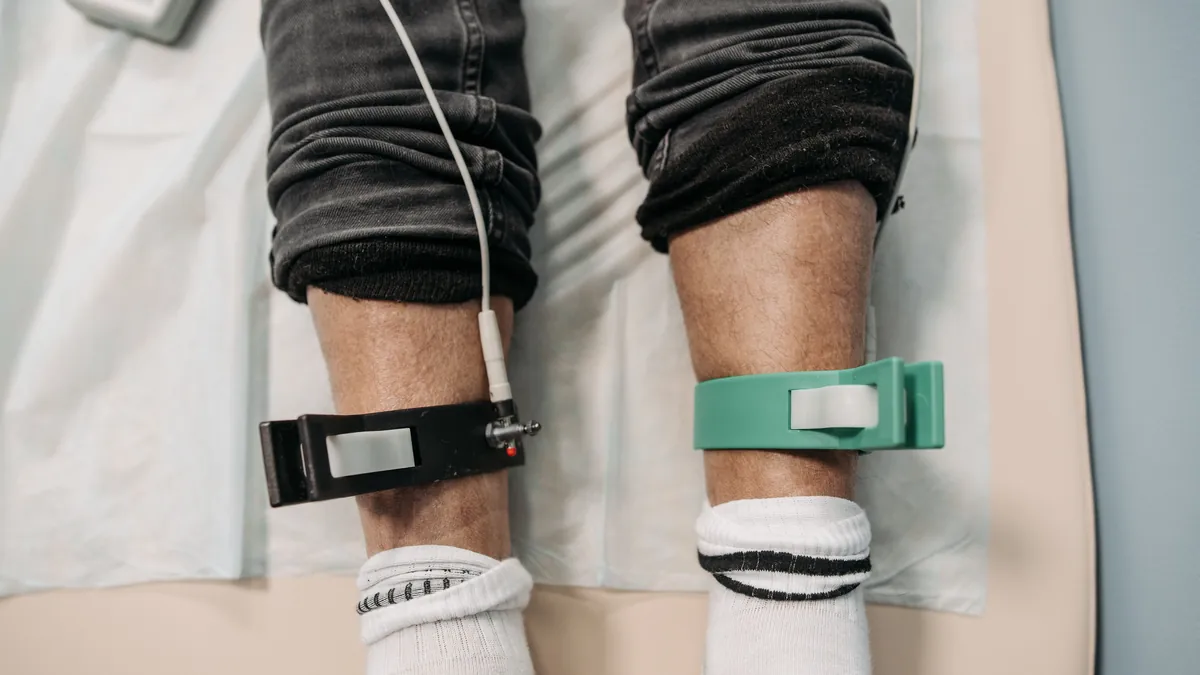
  • Electrode clips on a patient's lower legs during an in-office test

    Small-Fiber Versus Large-Fiber Neuropathy

    Small-fiber versus large-fiber neuropathy

    Small-fiber neuropathy damages the thin unmyelinated nerves that carry pain, temperature and sweating, and these fibers are typically affected first; large-fiber neuropathy damages the myelinated nerves that carry vibration, position sense, strength and reflexes. Nerve conduction studies measure the large fibers, while small fibers are measured with sudomotor testing, sensory testing or skin biopsy.

    If you have burning feet and a normal nerve conduction study, you were probably tested with the wrong instrument rather than reassured.

    What is the difference between small and large fiber neuropathy?

    Peripheral nerves come in two broad classes. Large myelinated fibers carry vibration, position sense, muscle power and reflexes. Small unmyelinated fibers carry pain, temperature, sweating and the control of blood vessels in the skin.

    Large myelinated fibersSmall unmyelinated fibers
    CarryVibration, position sense, strength, reflexesPain, temperature, sweating, skin blood flow
    Measured byNerve conduction studies, electromyographySudomotor testing, quantitative sensory testing, skin biopsy
    Typically affectedLaterEarlier
    SymptomsNumbness, weakness, unsteadiness, lost reflexesBurning, tingling, temperature loss, sweat changes
    Why a normal nerve conduction study does not exclude a neuropathy.

    Why are small nerve fibers affected first?

    They are long, thin and unmyelinated, which makes them metabolically expensive to maintain and dependent on a microcirculation that is itself vulnerable. Nerve injury associated with impaired glucose tolerance and metabolic syndrome preferentially affects them, and nerve conduction studies are relatively insensitive to that injury — which is why intraepidermal nerve fiber density on skin biopsy has traditionally been used to confirm the diagnosis.

    Can you have neuropathy with a normal nerve conduction study?

    A patient describes burning feet, worse at night. A nerve conduction study is ordered and comes back normal. The patient is told the nerves are fine. Two things have gone wrong: the instrument used cannot see the fiber population most likely to be affected, and the patient leaves believing nobody believes them.

    The American Diabetes Association’s position statement on diabetic neuropathy notes that a substantial proportion of distal symmetric polyneuropathy is painless — so the reverse trap also exists. The people at highest risk of a foot injury are frequently the ones reporting the fewest symptoms.

    How is small fiber neuropathy tested without a biopsy?

    Electrochemical skin conductance measures sweat gland function, which is driven by small unmyelinated sympathetic cholinergic fibers. It takes about three minutes, uses a very low voltage, and requires nothing more than placing palms and soles on metal plates. A systematic review appraised the method against established reference tests including the quantitative sudomotor axon reflex test, sympathetic skin responses, the thermoregulatory sweat test and skin biopsy.

    What does an abnormal small fiber test mean?

    An abnormal small-fiber result is a reason to look for a driver — glucose intolerance, B12 deficiency, thyroid disease, alcohol, medication, autoimmune disease — and a reason to protect the feet, particularly if sensation is also reduced. It is not a diagnosis by itself. Peripheral neuropathy symptoms.

    Frequently asked questions

    What are the symptoms of large fiber neuropathy?

    Large fiber neuropathy damages the myelinated nerves that carry vibration, position sense, strength and reflexes. The typical symptoms are numbness, weakness, unsteadiness and lost reflexes. These fibers are usually affected later than the small fibers, and they are the ones a nerve conduction study or electromyography measures.

    What are the symptoms of small fiber neuropathy in the feet?

    Small fiber neuropathy typically shows up as burning, tingling, loss of temperature sensation and changes in sweating, and burning feet are often worse at night. Some people have few or no symptoms at all, and they can be the ones at highest risk of a foot injury, because the usual warning signs are missing.

    What test confirms small fiber neuropathy?

    Skin biopsy measuring intraepidermal nerve fiber density has traditionally been used to confirm it. Small fibers can also be measured without a biopsy: sudomotor testing checks sweat gland function in about three minutes with your palms and soles resting on metal plates, and quantitative sensory testing is another option. A standard nerve conduction study cannot see these fibers.

    References

    • Cortez M, Singleton JR, Smith AG. Glucose intolerance, metabolic syndrome, and neuropathy. Handbook of Clinical Neurology. 2014;126:109–122. doi:10.1016/B978-0-444-53480-4.00009-6
    • Pop-Busui R, Boulton AJM, Feldman EL, et al. Diabetic Neuropathy: A Position Statement by the American Diabetes Association. Diabetes Care. 2017;40(1):136–154. doi:10.2337/dc16-2042
    • Novak P. Electrochemical skin conductance: a systematic review. Clinical Autonomic Research. 2019;29(1):17–29. doi:10.1007/s10286-017-0467-x

    Related reading