Category: Circulation

  • Healthcare worker measuring a patient's blood pressure with a cuff in a medical office.

    The Endothelial Glycocalyx: The Lining Sugar Strips First

    The Endothelial Glycocalyx: The Lining Sugar Strips First

    The endothelial glycocalyx is the soft gel lining the inside of your arteries that keeps blood cells from sticking, holds fluid inside the vessel and tells the artery when to widen.

    The inside of an artery is coated in a soft gel that decides how blood flows and what leaks. High blood sugar strips it in hours, long before a scan finds anything.

    Every artery you own is coated on the inside with a soft, sugar-based gel a fraction of a hair wide. The endothelial glycocalyx is that coating, and it does more work than its size suggests: it keeps blood cells from sticking, holds fluid and protein inside the vessel, and translates the push of flowing blood into the signal that tells an artery to widen. Nothing on a standard panel reports it, though vessel stiffness and dilation testing can measure the job it does. The reason it belongs in a conversation about blood sugar is timing. This layer is not lost over decades of plaque buildup. It thins in hours.

    A layer built to be sacrificed

    The glycocalyx is a wearable part. It is constantly built up and constantly shed, and that turnover is the point: a lining that can be stripped and replaced can absorb insult. Shedding is enzymatic, and the enzymes that cut it loose are switched on by oxidative stress and inflammatory signaling. When tearing down outruns building back, the cells underneath are left bare against flowing blood, and the layer that carried the widen-the-vessel signal is gone. Current vessel biology treats that imbalance in turnover as the root of what clinics call endothelial dysfunction. What follows is the ordinary sequence: fluid leaks where it should not, white cells and platelets find the wall sticky, and small arteries lose the ability to open on demand. Dr. Padda spent years telling people whose sugars sat a little above the line that this was not something to worry about yet. This is part of the physiology that changed his mind.

    What one high blood sugar does, measured

    The cleanest human demonstration is an old one. Investigators measured the whole-body volume of this layer in 10 healthy men by comparing how two tracers spread through the circulation, one able to enter the gel and one not. At rest the measurement repeated at about 1.7 liters. During a few hours of controlled high blood sugar, with insulin held steady so insulin itself was not the variable, it fell to roughly 0.8 liters. Hyaluronan, one of the layer’s own building blocks, rose in the blood as it came off the wall. Clotting markers rose with it, and vessel function worsened in the same session. An infusion that concentrated the blood without adding glucose did nothing, so this was the sugar, not the osmotic load.

    Read that against a glucose meter. A number that peaks after a meal and settles by morning is usually treated as a non-event because the fasting value looks fine. The lining does not average your day, and the silent inflammation sugar sets off runs on the same clock. The gums register the same post-meal injury in tissue you can see, discussed in what bleeding gums can say about sugar.

    From a spike to a pattern

    Measured in people who live with high blood sugar, the layer falls in steps. In adults with type 1 diabetes, glycocalyx volume ran about 1.5 liters in controls, lower in patients whose kidneys were not yet leaking protein, and about 0.2 liters in those with protein in the urine. The groups were tiny, 7 people each, so the direction matters more than the decimals: the layer is thinnest in the people whose smallest vessels have already declared themselves.

    In type 2 diabetes, the layer measured under the tongue and in the retina was thinner than in controls, and albumin escaped the capillaries faster. A newer sublingual camera found the same thinning in 65 adults with type 2 diabetes compared with 42 matched controls, alongside stiffer large arteries and changes in the retinal microcirculation. None of these measurements is a Measura test, and none is a routine clinical test anywhere; they are research tools, which is why the practical question becomes what a person can measure instead.

    Where your fat sits shows up in the lining

    The most useful recent finding has nothing to do with glucose numbers. Researchers measured a blood marker of shed glycocalyx in 219 adults without diabetes and 203 adults with type 2 diabetes, and compared it against imaging and body composition. Median levels ran 16.0 nanograms per milliliter in the group without diabetes and 26.5 in the group with it. Within each group the marker rose with the amount of fat packed inside the abdomen. Neither body mass index nor the fat under the skin tracked it at all. The difference between those compartments is laid out in visceral versus subcutaneous fat.

    That is the second biological driver, and it is not sugar. Visceral fat is an endocrine organ, and the metaflammation it pumps into the circulation is the switch that turns shedding enzymes on. Insulin resistance sits underneath both: it fills that depot and keeps post-meal glucose elevated. The third driver is not biology at all. Refined starch and industrial seed oils are engineered to be the most available calories on every shelf, so the post-meal spike is a feature of the food supply rather than a personal failure. A layer stripped several times a day, every day, never gets a quiet stretch to rebuild.

    What a standard visit sees, and what it skips

    A routine visit takes a blood pressure at the arm and orders a cholesterol panel and perhaps an A1C. All three are worth having, and all three read late. An A1C is a three-month average that hides the spikes doing the damage, while a fasting insulin result reads the compensation years earlier. A cholesterol panel counts particles; it says nothing about whether your vessels can open. Blood pressure at the arm is a downstream consequence of stiffness that has already set in. The checkup measures the smoke after the fire has spread, then calls a normal reading reassurance.

    What Measura can measure in the same person

    Measura [Cardiometabolic and Autonomic Health Analysis] is a testing service. It measures and sends findings to your physician; it does not treat, and it cannot image your glycocalyx. What it can do is measure the functions that layer is responsible for, in you, today.

    • Arterial stiffness and endothelial function testing asks how stiff your larger arteries are and how well the lining lets them widen, the job the glycocalyx starts.
    • Photoplethysmography uses light at the skin to read the pulse in the smallest vessels, where leaking and sticking happen first.
    • Pulse volume recording traces the pulse waveform along the arms and legs to show how blood is moving through those arteries.
    • Laboratory panels cover the drivers: fasting insulin reported beside glucose, inflammatory markers, and lipid particle detail rather than total cholesterol alone.

    A single reading is a starting point, not a verdict. What endothelial dysfunction is and how it is tested covers the vessel-function side once results are in hand, and why a normal ankle-brachial index can be misleading explains how a reassuring circulation number can still miss small-vessel trouble.

    What appears to move it

    Human intervention evidence is thin, because the trials that would settle it isolate one disease and exclude the comorbid metabolic patient described here. What exists points one way. In fifty healthy young adults doing sprint-interval training, a thicker measured glycocalyx went with greater exercise capacity both before and after the program. In adults with type 2 diabetes given a glycocalyx building block by mouth for 2 months, the measured layer thickened and albumin leak trended back toward normal. In 28 adults with type 2 diabetes and 33 without who had bariatric surgery, shedding markers improved over two years, and what best predicted the improvement was loss of body weight and fat mass and a gain in insulin sensitivity.

    Put plainly, the layer responds to the terrain, not to a single number. Lowering the height of your after-meal glucose, shrinking the visceral depot, and moving hard enough to make your circulation work are the levers with mechanism behind them. Ask your physician for fasting insulin next to glucose, ask what your after-meal numbers look like rather than the average, and ask whether a vessel-function measurement belongs beside the arm cuff. Finding cardiovascular risk early describes how that testing fits a checkup. Medication decisions stay with your physician, never with a single result. Unmeasured is unmanaged, and this is a layer nobody has ever measured in you. When glucose delivery to the brain falters, a second fuel can still get in, the subject of the fuel an insulin-resistant brain can still use.

    Frequently asked questions

    Can my glycocalyx be tested at my appointment?

    Not at Measura, and not in routine care anywhere. The research above used tracer dilution, a sublingual microscope camera or retinal angiography, all research methods. What can be measured in the same person is the function that layer governs: arterial stiffness and dilation, small-vessel pulse, and the metabolic blood work behind both. What a test result can and cannot tell you sets the expectation.

    Does a normal A1C mean my vessel lining is safe?

    An A1C is an average across about three months, and the damage documented here followed hours of raised glucose. A person with an unremarkable average can still spike hard after meals, and the layer responds to the peak, not the mean. Insulin usually rises years before glucose does, which is why it belongs on the panel. High insulin with normal blood sugar explains that gap.

    Why does my waist matter more than my weight here?

    Because the blood marker of a shed lining tracked the fat packed inside the abdomen and tracked neither body mass index nor the fat under the skin. That depot sends inflammatory signals into the circulation that switch on the enzymes stripping the layer, so two people at the same weight can be in different vascular situations. How to measure visceral fat covers what separates the two.

    If I cannot measure the layer, what should I track?

    Track what drives it and what it serves: fasting insulin beside glucose, a marker of inflammation, where your fat sits, and a vessel-function measurement you can repeat under the same conditions. One set of numbers now becomes the baseline that tells you, a year from now, whether anything you changed reached your arteries. What insulin resistance looks like before diabetes describes the earliest stage.

    Measure what the lining protects

    Request Measura testing for vessel function, small-vessel pulse and metabolic blood work, with results sent to your physician. Bring the report to the clinician who manages your blood sugar.

    4477 Woodson Rd, Suite 201, St. Louis, MO 63134. Monday to Friday, 9:00 a.m. to 5:00 p.m. Please do not send symptoms, diagnoses or images through a web form — a website form is not a secure medical channel. Send your name and number and we will call you back.

    References

    • Nieuwdorp, M., van Haeften, T. W., Gouverneur, M. C., Mooij, H. L., van Lieshout, M. H., Levi, M., Meijers, J. C., Holleman, F., Hoekstra, J. B., Vink, H., Kastelein, J. J., & Stroes, E. S. (2006). Loss of endothelial glycocalyx during acute hyperglycemia coincides with endothelial dysfunction and coagulation activation in vivo. Diabetes, 55(2), 480-486. https://doi.org/10.2337/diabetes.55.02.06.db05-1103
    • Nieuwdorp, M., Mooij, H. L., Kroon, J., Atasever, B., Spaan, J. A., Ince, C., Holleman, F., Diamant, M., Heine, R. J., Hoekstra, J. B., Kastelein, J. J., Stroes, E. S., & Vink, H. (2006). Endothelial glycocalyx damage coincides with microalbuminuria in type 1 diabetes. Diabetes, 55(4), 1127-1132. https://doi.org/10.2337/diabetes.55.04.06.db05-1619
    • Broekhuizen, L. N., Lemkes, B. A., Mooij, H. L., Meuwese, M. C., Verberne, H., Holleman, F., Schlingemann, R. O., Nieuwdorp, M., Stroes, E. S., & Vink, H. (2010). Effect of sulodexide on endothelial glycocalyx and vascular permeability in patients with type 2 diabetes mellitus. Diabetologia, 53(12), 2646-2655. https://doi.org/10.1007/s00125-010-1910-x
    • Miyamoto, S., Kakutani, Y., Morioka, T., Yamazaki, Y., Ochi, A., Fukumoto, S., Shoji, T., & Emoto, M. (2025). Association of Visceral Fat Accumulation with Endothelial Glycocalyx Degradation in People with and without Type 2 Diabetes: A Retrospective Cross-sectional Study. Journal of Atherosclerosis and Thrombosis, 32(12), 1558-1570. https://doi.org/10.5551/jat.65623
    • Agapitou, C., Sergentanis, T. N., Thymis, J., Pavlidis, G., Lampsas, S., Korakas, E., Kountouri, A., Pliouta, L., Karmiris, E., Lagiou, A., Theodossiadis, P., Lambadiari, V., Ikonomidis, I., & Chatziralli, I. (2024). Retinal Microvascular Changes in Association with Endothelial Glycocalyx Damage and Arterial Stiffness in Patients with Diabetes Mellitus Type 2: A Cross-Sectional Study in a Greek Population. Journal of Personalized Medicine, 14(9), 995. https://doi.org/10.3390/jpm14090995
    • Andersson, M., Agren, A., Henriksson, P., Wallen, H., & Thorell, A. (2025). Influence of Bariatric Surgery on Endothelial and Glycocalyx Biomarkers in Obesity and Type 2 Diabetes. The Journal of Clinical Endocrinology and Metabolism, 110(10), e3193-e3202. https://doi.org/10.1210/clinem/dgaf092
    • Schmitz, B., Niehues, H., Lenders, M., Thorwesten, L., Klose, A., Kruger, M., Brand, E., & Brand, S. M. (2019). Effects of high-intensity interval training on microvascular glycocalyx and associated microRNAs. American Journal of Physiology. Heart and Circulatory Physiology, 316(6), H1538-H1551. https://doi.org/10.1152/ajpheart.00751.2018
    • Gomez Toledo, A., Golden, G. J., Cummings, R. D., Malmstrom, J., & Esko, J. D. (2025). Endothelial Glycocalyx Turnover in Vascular Health and Disease: Rethinking Endothelial Dysfunction. Annual Review of Biochemistry, 94(1), 561-586. https://doi.org/10.1146/annurev-biochem-032620-104745


    Related reading

  • Dr. Gurpreet Singh Padda beside the Chapter 9 title card of The Angry Gut reading Your Gut Is Suffocating.

    What Is Endothelial Dysfunction? The Warning Your Arteries Send

    Your Gut Is Suffocating | The Angry Gut, Chapter 9

    What Is Endothelial Dysfunction? The Warning Your Arteries Send

    Endothelial dysfunction is the failure of the blood vessel lining to make enough nitric oxide to widen small arteries when tissue needs more blood. It shows up as pain after meals, rising blood pressure or erection problems, and it is tested by measuring arterial stiffness and endothelial function, not by a cholesterol panel.

    Your blood vessels have a lining that decides, moment to moment, whether they can open. When it falters, the first complaints often come from organs nobody thinks to connect.

    What is endothelial dysfunction? It is the failure of the thin lining inside your blood vessels, the endothelium, to make enough nitric oxide to let small arteries widen when a tissue needs more blood. It rarely announces itself as a heart problem. It tends to show up as separate complaints that get sent to separate specialists. The video above, Your Gut Is Suffocating, tells that story from Chapter 9 of The Angry Gut by Dr. Gurpreet Singh Padda, MD, MBA, MHP, and Ami Michelle Grimes. Here the focus is measurement: what a routine visit checks, what it leaves out, and which tests show how your vessels actually behave.

    What is endothelial dysfunction? One lining, many complaints

    Eating is a demand on your circulation. Blood flow to the bowel rises by 30-150% after a meal, so an artery that manages fine while you sit still may not keep up with lunch. An erection is a nitric oxide event in a small artery. Blood pressure depends on how easily vessels relax. When the lining falters, each of those can fail on its own schedule and be handled on its own: pain after lunch goes to gastroenterology, erection problems to urology, blood pressure to cardiology. Three offices, one vessel lining.

    Your gut is the first brain, and the second brain sits in your skull. The first brain is fed from behind its lining, and the tips of the tiny absorbing fronds called villi sit at the far end of that supply line. When blood runs short, that is where the shortfall lands first, and tissue that is not getting blood cannot heal well.

    What causes endothelial dysfunction?

    Two biological routes matter most. The enzyme that makes nitric oxide needs raw materials, and when they run low it uncouples and produces superoxide, a corrosive radical, so the machinery built to open vessels starts damaging them. Oxidized LDL, cholesterol-carrying particles that have gone rancid, binds a receptor on the vessel lining and sets off the same chemistry. In 4,658 people followed for nearly twenty years, those with the highest circulating levels of that receptor had 1.76 times the risk of a first heart attack.

    There is also a backup route most people never hear about. Nitrate from green leafy vegetables is concentrated into saliva, and bacteria at the back of your tongue turn it into nitrite, which the body can use to make nitric oxide. When 19 healthy volunteers used antiseptic mouthwash for seven days, nitrite production in the mouth dropped by 90% and blood pressure rose by 2-3.5 mmHg. In one cohort, rinsing twice a day or more went with 1.85 times the rate of new high blood pressure. The effect shows up mainly in older adults and people already treated for hypertension, whose main enzyme is already struggling.

    Then there is the economic driver. Frying oil is cheapest at the end of its life, reused in fryers that are changed least often, and the people with the fewest food choices eat the most of it. The blood tests behind reassuring vegetable oil studies read an intact fatty acid, drawn once, and cannot see what oxidized before it was swallowed. Are seed oils bad for you covers that measurement gap.

    Does a routine checkup test for endothelial dysfunction?

    A standard visit measures blood pressure at the arm and orders a cholesterol panel. Both are useful, and neither tells you whether your vessel lining can open on demand. A scope looks at the inside surface of the gut and cannot see the arteries feeding it from behind. The man at the center of the video had four scopes before anyone imaged an artery. He also had a normal body weight, and the European guideline now names the expectation of a thin, wasted patient as a pitfall that delays diagnosis.

    Erection problems are the most overlooked signal of all. Pooling 14 studies with 92,757 men, erectile dysfunction predicted heart attack at 1.62 times the usual risk. Among men with coronary disease who had both conditions, the sexual symptom came first in 93% of them, by about two years. It is usually answered with a prescription and no further questions. The medicine works. The silence after it is the problem.

    How is endothelial dysfunction tested?

    Measura [Cardiometabolic and Autonomic Health Analysis] does not image the arteries that supply your intestines. If meals reliably bring pain, or you have started eating less to avoid it, imaging of those mesenteric arteries is a separate test done elsewhere, and your physician needs to hear about the pattern. What Measura can show is how the vessels you share with the rest of your body are behaving:

    • Arterial stiffness and endothelial function measures how stiff your larger arteries are and how well the vessel lining lets them widen.
    • Pulse volume recording records the pulse waveform at several points along your arms and legs to show how blood moves through those arteries.
    • Ankle-brachial index sets the pressure reading taken at your ankle against the one taken at your arm, a simple screen for narrowing in the legs.
    • Laboratory panels cover blood work on glucose, insulin and lipids, the metabolic conditions that wear on the vessel lining.

    A normal result does not rule out a narrowed gut artery, and an abnormal one does not diagnose a disease by itself. Findings go to your physician, who decides what needs imaging or treatment. Why a normal ankle-brachial index can be misleading explains one of those limits.

    What to ask for

    • If pain reliably follows meals, ask whether anyone has looked at the arteries, not just the lining of the gut.
    • If you have erection problems, tell your primary care physician, not only the doctor who wrote the prescription.
    • If you use antiseptic mouthwash twice a day, ask your dentist what it is treating and when the course should end.
    • If your blood pressure keeps climbing, ask whether a vascular measurement would add to the arm cuff reading.

    Do not change any medication on your own; bring these questions to the physician who prescribed it. Dr. Padda was trained, like every physician, to send each complaint to the organ that reports it, and the humbling lesson of this case is that the gut could not be rescued from inside the gut. The Chapter 9 book companion page carries every study with its full numbers, and finding cardiovascular risk early explains how vascular testing fits a checkup. Before the vessel wall itself stiffens, high glucose strips the thin sugar coat that lines it, described in the endothelial glycocalyx.

    Frequently asked questions

    What are the symptoms of endothelial dysfunction?

    There is no single symptom. The lining fails quietly, and the first complaints come from whichever tissue has the least reserve: pain after meals, erection problems, blood pressure that keeps rising, or legs that tire when you walk. Because each goes to a different specialist, the shared cause is easy to miss. Leg symptoms and circulation covers the limb side.

    Can mouthwash raise blood pressure?

    An antiseptic rinse wipes out the bacteria on the back of your tongue that convert nitrate from vegetables into nitrite. Among healthy volunteers, seven days of rinsing cut oral nitrite production by 90% and raised blood pressure by a few points, and twice-daily use tracked with new hypertension in a cohort. A short course for a dental problem is different from years of habit. Questions worth asking your doctor helps you raise it.

    Is erectile dysfunction a sign of heart disease?

    It often comes first. Pooled studies link erectile dysfunction to later heart attacks, and among men already diagnosed with coronary disease, most noticed the erection problem roughly two years before any chest symptoms. It reflects how well small arteries open, which is why it deserves a cardiovascular question and not only a prescription. Who should have cardiometabolic testing explains who benefits.

    Can Measura find a blocked artery to my intestines?

    No. Measura does not image the mesenteric arteries that feed the gut; that is a separate imaging test your physician can arrange. Measura measures how your larger arteries stiffen and open, how blood moves through your limbs, and the metabolic blood work behind vessel health, which describes the terrain your gut shares with the rest of you. What to expect at your appointment describes the visit.

    Does a normal cholesterol panel mean my arteries are healthy?

    Not necessarily. A cholesterol panel describes fat-carrying particles in your blood, not whether your vessel lining can widen when tissue needs more blood. People with normal cholesterol can still have stiff arteries or a struggling endothelium, especially with high insulin or rising blood pressure. Measuring the vessel directly answers a different question. What insulin resistance looks like before diabetes explains one hidden driver.

    What damages the lining of your blood vessels?

    Two routes matter most. When the enzyme that makes nitric oxide runs short of raw materials, it produces superoxide, a corrosive radical, so the machinery built to open vessels starts damaging them. Oxidized LDL, cholesterol-carrying particles that have gone rancid, binds a receptor on the lining and sets off the same chemistry. Reused frying oil is the economic driver: cheapest, eaten most where food choices are fewest, and invisible to standard blood tests.

    What foods help your blood vessels make nitric oxide?

    Green leafy vegetables supply a backup route. Their nitrate is concentrated into saliva, and bacteria at the back of your tongue turn it into nitrite, which the body can use to make nitric oxide. That route matters most for older adults and people already treated for high blood pressure. Antiseptic mouthwash wipes out those bacteria, so a twice-daily rinse habit can undercut the vegetables on your plate.

    Find out how your vessels open

    Request Measura testing to see how your arteries stiffen and dilate, and bring the results to the physician who knows your symptoms.

    4477 Woodson Rd, Suite 201, St. Louis, MO 63134. Monday to Friday, 9:00 a.m. to 5:00 p.m. Please do not send symptoms, diagnoses or images through a web form — a website form is not a secure medical channel. Send your name and number and we will call you back.

    References

    • Terlouw, L. G., Moelker, A., Abrahamsen, J., Acosta, S., Bakker, O. J., Baumgartner, I., Boyer, L., Corcos, O., van Dijk, L. J., Duran, M., Geelkerken, R. H., Illuminati, G., Jackson, R. W., Kärkkäinen, J. M., Kolkman, J. J., Lönn, L., Mazzei, M. A., Nuzzo, A., Pecoraro, F., … Bruno, M. J. (2020). European guidelines on chronic mesenteric ischaemia – joint United European Gastroenterology, European Association for Gastroenterology, Endoscopy and Nutrition, European Society of Gastrointestinal and Abdominal Radiology, Netherlands Association of Hepatogastroenterologists, Hellenic Society of Gastroenterology, Cardiovascular and Interventional Radiological Society of Europe, and Dutch Mesenteric Ischemia Study group clinical guidelines on the diagnosis and treatment of patients with chronic mesenteric ischaemia. United European Gastroenterology Journal, 8(4), 371-395. https://doi.org/10.1177/2050640620916681
    • Janaszak-Jasiecka, A., Siekierzycka, A., Płoska, A., Dobrucki, I. T., & Kalinowski, L. (2021). Endothelial dysfunction driven by hypoxia—The influence of oxygen deficiency on NO bioavailability. Biomolecules, 11(7), 982. https://doi.org/10.3390/biom11070982
    • Schiopu, A., Björkbacka, H., Narasimhan, G., Loong, B. J., Engström, G., Melander, O., Orho-Melander, M., & Nilsson, J. (2023). Elevated soluble LOX-1 predicts risk of first-time myocardial infarction. Annals of Medicine, 55(2), 2296552. https://doi.org/10.1080/07853890.2023.2296552
    • Kapil, V., Haydar, S. M., Pearl, V., Lundberg, J. O., Weitzberg, E., & Ahluwalia, A. (2013). Physiological role for nitrate-reducing oral bacteria in blood pressure control. Free Radical Biology & Medicine, 55, 93-100. https://doi.org/10.1016/j.freeradbiomed.2012.11.013
    • Joshipura, K., Muñoz-Torres, F., Fernández-Santiago, J., Patel, R. P., & Lopez-Candales, A. (2020). Over-the-counter mouthwash use, nitric oxide and hypertension risk. Blood Pressure, 29(2), 103-112. https://doi.org/10.1080/08037051.2019.1680270
    • Vlachopoulos, C. V., Terentes-Printzios, D. G., Ioakeimidis, N. K., Aznaouridis, K. A., & Stefanadis, C. I. (2013). Prediction of cardiovascular events and all-cause mortality with erectile dysfunction: A systematic review and meta-analysis of cohort studies. Circulation: Cardiovascular Quality and Outcomes, 6(1), 99-109. https://doi.org/10.1161/CIRCOUTCOMES.112.966903
    • Montorsi, P., Ravagnani, P. M., Galli, S., Rotatori, F., Veglia, F., Briganti, A., Salonia, A., Dehò, F., Rigatti, P., Montorsi, F., & Fiorentini, C. (2006). Association between erectile dysfunction and coronary artery disease. Role of coronary clinical presentation and extent of coronary vessels involvement: The COBRA trial. European Heart Journal, 27(22), 2632-2639. https://doi.org/10.1093/eurheartj/ehl142
    • Marklund, M., Wu, J. H. Y., Imamura, F., Del Gobbo, L. C., Fretts, A., de Goede, J., Shi, P., Tintle, N., Wennberg, M., Aslibekyan, S., Chen, T. A., de Oliveira Otto, M. C., Hirakawa, Y., Eriksen, H. H., Kröger, J., Laguzzi, F., Lankinen, M., Murphy, R. A., Prem, K., … Risérus, U. (2019). Biomarkers of dietary omega-6 fatty acids and incident cardiovascular disease and mortality: An individual-level pooled analysis of 30 cohort studies. Circulation, 139(21), 2422-2436. https://doi.org/10.1161/CIRCULATIONAHA.118.038908

    Related reading

  • A blood pressure cuff and stethoscope on a patient's upper arm, one half of the ankle-brachial index comparison

    Why a Normal Ankle-Brachial Index Can Be Misleading

    Why a normal ankle-brachial index can mislead

    A normal ankle-brachial index can be misleading when the ankle arteries are calcified, as they often are in long-standing diabetes and chronic kidney disease. The stiff artery resists the cuff, so the ankle pressure reads falsely high and the ratio looks normal. The toe-brachial index and pulse volume recording are measured instead.

    A calcified artery cannot be squeezed. The cuff reads high, the ratio looks fine, and a leg with real disease produces a reassuring number.

    How is the ankle-brachial index calculated?

    The ankle-brachial index is the highest systolic pressure measured at the ankle divided by the highest measured at the arm. In a healthy circulation the two are close, so the ratio sits near one. When an artery upstream is narrowed, ankle pressure falls and the ratio drops.

    It is non-invasive, takes minutes, and is the standard first measurement for lower-limb arterial disease. Its measurement and interpretation are set out in detail in the American Heart Association scientific statement on the subject.

    Why can a normal ankle-brachial index be wrong?

    The measurement depends on the cuff being able to compress the artery. Medial calcification — calcium in the vessel wall — makes the artery stiff and resistant to compression. The cuff has to be inflated to a higher pressure before flow stops, so the recorded ankle pressure is falsely high and the ratio comes out normal or even elevated.

    The populations in which this happens are exactly the populations being screened: long-standing diabetes and chronic kidney disease in particular.

    What does a high ankle-brachial index mean?

    An index that is unexpectedly high is not a healthier artery. It is usually a stiffer one, and it should be treated as an uninterpretable result rather than a good one.

    What test is used when the ankle-brachial index is unreliable?

    • The toe-brachial index. The small digital arteries in the toes are much less prone to medial calcification, so a toe measurement stays valid where the ankle one does not.
    • Pulse volume recording. The waveform reflects the volume change in the limb with each heartbeat and does not depend on compressing the artery at all. Because Doppler sensitivity is itself reduced by calcification, volume plethysmography is often the practical way to obtain a peripheral waveform in these patients.
    • Pulse waveform contour at the toe. Measured optically, and reflecting flow at the far end of the circulation.

    How do I interpret my ABI results?

    IndexWaveformReading
    ReducedDampedConsistent with significant arterial disease
    Normal or elevatedDampedSuspect calcification — weight the waveform and the toe index
    NormalNormalAgainst a significant arterial cause; look elsewhere for the symptom
    Markedly asymmetricAsymmetricAsymmetric disease, clinically meaningful even within range
    Why all three measurements are taken in the same study.

    The practical consequence

    If you have diabetes or kidney disease and have been told your circulation is fine on the basis of an ankle pressure alone, that is worth revisiting — particularly if you have symptoms, a slow-healing sore, or feet that have stopped sweating. Leg symptoms and circulation.

    Frequently asked questions

    What are normal ABI readings?

    In a healthy circulation the pressure at the ankle and the pressure at the arm are close, so the ratio sits near one. When an artery upstream is narrowed, ankle pressure falls and the ratio drops. A reading that looks normal or even high is only reassuring if the artery can be compressed, which is not the case when its wall is calcified.

    Can you have peripheral artery disease with a normal ABI?

    Yes. Medial calcification, calcium in the artery wall, makes the vessel stiff and resistant to the cuff, so the recorded ankle pressure is falsely high and the ratio comes out normal. This happens most in long-standing diabetes and chronic kidney disease, the same people most often screened. A damped waveform alongside a normal index is the clue.

    What is the toe-brachial index?

    It compares blood pressure in the toe with pressure in the arm. The small arteries in the toes are much less prone to medial calcification than the ankle arteries, so a toe measurement stays valid where the ankle one does not. It is taken in the same study as the ankle-brachial index and the pulse volume waveform.

    References

    • Aboyans V, Criqui MH, Abraham P, et al. Measurement and interpretation of the ankle-brachial index: a scientific statement from the American Heart Association. Circulation. 2012;126(24):2890–2909. doi:10.1161/CIR.0b013e318276fbcb
    • Fowkes FGR, Rudan D, Rudan I, et al. Comparison of global estimates of prevalence and risk factors for peripheral artery disease in 2000 and 2010: a systematic review and analysis. Lancet. 2013;382(9901):1329–1340. doi:10.1016/S0140-6736(13)61249-0

    Related reading

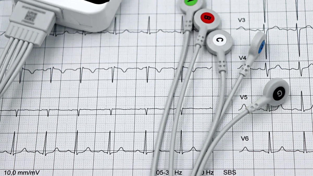
  • Close-up of ECG monitor leads on a printed cardiogram.

    Leg Pain When You Walk: Claudication Explained

    Leg pain when you walk

    Leg pain when you walk that eases once you stand still is usually claudication, the classic sign of peripheral artery disease: arteries narrowed by plaque cannot raise blood flow to working muscle. The ankle-brachial index measures it in minutes.

    Cramping that appears at a predictable distance and disappears within minutes of stopping is one of the few symptoms in medicine with a simple, objective test behind it.

    What causes claudication?

    Peripheral artery disease is the narrowing of arteries by atherosclerotic plaque along the vessel lining. As plaque accumulates the artery narrows, stiffens and loses function, and blood flow falls — usually to the legs first.

    Muscle that is working needs more blood than muscle at rest. When the supply cannot rise to meet demand, the muscle produces a cramping, aching or tired sensation that stops when the demand stops. Hence the pattern: predictable onset distance, relief within minutes of standing still, and reproducibility from one day to the next.

    What can be mistaken for claudication?

    PatternMore likely
    Cramping at a predictable walking distance, relieved by standing stillArterial claudication
    Aching and heaviness after standing still for a long time, relieved by elevationVenous
    Pain radiating from the back down the leg, worse with certain positions, relieved by leaning forwardSpinal or nerve root
    Burning or tingling at rest, worse at nightSmall-fiber neuropathy — see burning feet at night
    Pain in the foot at night relieved by hanging it out of bedMore advanced ischemia — needs prompt assessment
    Leg symptom patterns. None of these is a diagnosis on its own.

    Why do so many people have artery disease without knowing it?

    Because people adapt. Walking distance shortens quietly, a hill gets avoided, a parking space closer to the door gets chosen, and the symptom never becomes a complaint. Peripheral artery disease is the third leading cause of atherosclerotic cardiovascular morbidity after coronary disease and stroke, and an estimated 202 million people were living with it worldwide in 2010.

    Prevalence rises steeply with age — from roughly 5% of adults in their late forties to roughly 18% by the late eighties in the wealthier countries surveyed. The strongest associations were current smoking, then diabetes, hypertension and high cholesterol.

    How is claudication tested?

    The ankle-brachial index compares systolic pressure at the ankle with the arm and takes minutes. Because a calcified artery can make that index falsely reassuring, it is paired with the pulse volume waveform and the toe-brachial index, neither of which depends on the vessel being compressible. Why that matters.

    Why find peripheral artery disease early?

    Not primarily because of the legs. The same process is happening in the coronary and cerebral circulation, and a measurable abnormality in an accessible artery is information about all of them. It is also one of the areas where the interventions — stopping smoking, supervised walking, treating blood pressure and glucose — are well established and measurable.

    Frequently asked questions

    What does claudication feel like in the legs?

    It feels like cramping, aching or tiredness in working leg muscle that shows up at a predictable walking distance and stops within minutes of standing still. The pattern repeats from one day to the next. Pain that radiates from the back and eases when you lean forward, or burning and tingling at rest, points to other causes.

    How long does it take for claudication to go away?

    An episode of claudication eases within minutes of standing still, because the muscle stops demanding more blood than narrowed arteries can deliver. That quick relief at rest, together with a predictable onset distance, is what separates it from venous aching, which eases with elevation, and from spinal pain, which eases by leaning forward.

    How do you fix claudication?

    Claudication is one of the areas where the interventions are well established and measurable: stopping smoking, supervised walking, and treating blood pressure and glucose. The first step is to confirm it. The ankle-brachial index takes minutes, and the pulse volume waveform and toe-brachial index cover the legs where calcified arteries make the index read falsely normal.

    References

    • Fowkes FGR, Rudan D, Rudan I, et al. Comparison of global estimates of prevalence and risk factors for peripheral artery disease in 2000 and 2010: a systematic review and analysis. Lancet. 2013;382(9901):1329–1340. doi:10.1016/S0140-6736(13)61249-0
    • Aboyans V, Criqui MH, Abraham P, et al. Measurement and interpretation of the ankle-brachial index: a scientific statement from the American Heart Association. Circulation. 2012;126(24):2890–2909. doi:10.1161/CIR.0b013e318276fbcb

    Related reading